juliana

Page 132

image

image

Figure 2–59. Electron micrographs of nuclei. 
A — nucleus in a cell of mouse pancreas (After De Robertis at al., 1973). 
1 — chromatin connected with the nucleolus (2); 3 — nucleoplasm; the 
arrows indicate pores in the nuclear envelope. 
B — nucleus in a cell of the dividing area in a root of Zea mays. Bar 0.8 μm 
(Courtesy of S. Doncheva and G. Ignatov, Institute of Plant Physiology, 
Sofia). 
NE — nuclear envelope; N — nucleus; n — nucleolus.

The nucleus is filled up with nucleoplasm limited by a nuclear envelope,
consisting of two membranes — inner and outer. They are separated by a
intermediate space. The nuclear envelope is crossed by pores of diameter
10—20 nm through which nucleic acids, proteins and various other

Page 132 Read More »

Page 131

image

Figure 2–58. Three-dimensional view of a small section of a cell 
membrane (After Alberts et al., 1989)

Specialized protein complexes are located in the liquid layer of the lipid
membranes. The lipoproteins are in the lipid phase and are fixed through
hydrophobic bonds (integral proteins). Hydrophilic proteins are connected
by means of electrostatic bonds with inner and outer surface of the
membrane and interact with hydrophilic heads of the polar lipids. The main
role in the membrane formation is played by the hydrophilic bonds: lipid—
lipid, lipid—protein, protein—protein. The thickness of plasmalemma does
not exceed 6—10 nm.
Proteins also participate in plasmalemma. They act as enzymes,
pumps, carriers, ionic channels, and also as regulator proteins and
structural proteins. The labile structure of the cell membrane enables it to
perform different functions — barrier, transport, osmotic, electric, energetic,
biosynthetic, secretory, receptor-regulating, etc.

Nucleus

Cell nucleus is an obligatory and irreplaceable constitutive organelle of
eukaryotic cells. Its shape can be different — spheric, elliptic, prolonged,
leaf-like, etc. Sometimes it follows the form of cell. To considerable extent
the size of nucleus depends on the size of cell, its functions and
specialization. In lower unicellular eukaryotes it can be of the order of 1—2
μm, and in higher organisms varies from 4 to 40 μm. Nuclei of animal and
plant cell are shown in Figure 2–59 A, B.

Page 131 Read More »

Page 130

There are shapely cell walls in independently existed unicellular
organisms (bacteria, paramecia, infusoria, euglena, etc.), as well as in some
cells of the higher plant and animal organisms. A clear cytological picture of
cell wall, plasmalemma and plasmodesma can to be seen in Figure 2–56.

image

Figure 2–56. Cell wall, plasmalemma and plasmodesma in a root of 
Allium cepa (After Frey-Wissling and Mühlethaler, 1965)

image

Figure 2–57. Cross-section 
of plasmic membrane of 
human erythrocyte (From 
Yost, 1975).

In some cells (mainly animal) participating in tissues and organs of
multicellular organisms or strictly specialized, the outer cell wall is absent or it is not
shapely. Plasmic membrane of human erythrocyte, where outer cell wall is not
observed, is shown in Figure 2–57. At present, the liquid-mosaic hypothesis on
plasmic membrane structure is the most appreciated one. According this hypothesis the
membrane is built of a double lipid layer and some amount of protein molecules built-in. The
lipid molecules are facing with their hydrophobic ends. Diagrammatically this is shown in Figure
2–58. Besides, the membrane contains glycolipids, sterines, aliphatic acids, etc. The
lipids participating in the double membrane layer are labile compounds and their molecules
change the position and mutual location continuously.

Page 130 Read More »

Page 129

image

image

Figure 2–55. Cell wall of the sea alga Valonia (After Steward and 
Mühlethaler, 1953).
a — primary wall, with disperse texture; b — secondary wall, with 
crossed parallel texture.

Page 129 Read More »

Page 128

image

Figure 2–54 B. Pinocytosis in the cells of the root tip of Ricinus
communis (After Frey-Wissling and Mühlethaler, 1965).

The notion of the cell envelope has been altered in the course of
studying the cell. As it was already mentioned, for a long time it has been
considered usual barrage wall of no special importance. Later on it became
clear that it is a multilayer formation performing various biological functions.
The chemical composition and structural organization of the cell
envelope have been an object of numerous investigations prompted by its
importance for cell formation and functioning as a biological system, as well
as by the historical fact that it is the first structure seen by R. Hooke in 16 65.
It is the cause for the discovery of cell, thus making a start with its
studying. The basic components of cell envelope are different
polysaccarides (mainly cellulose and starch), proteins, lipids, etc. The water
content is high in it — up to 95%. In the course of binding single
polysaccaride chains there are formed threads gathered together in
fascicles and shaped like sticks. It was established that they possess
dispersed and crossed parallel texture (Fig. 2–55).
There have been long arguments about the presence or absence of
outer cell wall. The reasons for that are mainly two: on one hand,
morphologically it has not been clearly observed under light microscope,
and on the other — in some cases it really is absent. Contemporary
biochemical and physical methods, especially using the electron
microscopy, made it possible to establish that normally the cells possess a
surface layer (cell wall) of high-molecular carbohydrates or in a complex
with proteins, that performs locomotory and protective functions and
internal plasmic membrane (plasmalemma) built mainly of lipid and protein
molecules.

Page 128 Read More »

Page 127

registered by means of cytological methods (Fig. 2–54 B). The mechanisms
of including, transporting and excreting substances in the cells are not
enough elucidated on molecular level.

image

Figure 2–54 A. Schematic representation of the pinocytosis. 

Page 127 Read More »

Page 126

Depending on that, whether the basic genetic material (DNA) is
located “freely” in cytoplasm or in a well defined nucleus encircled by a
double membrane, the cells are divided into prokaryotic (protocists) and
eukaryotic (eucists). Bacteria and unicellular blue-green algae are typical
prokaryotic cells. All the other cells — either independently existing, or
included in the composition of multicellular organisms — are eukaryotic.
Irrespective of some differences and peculiarities the cells possess
similar internal organization. Each cell possesses an envelope, nucleus or
nuclear equivalent (nucleoid) and cytoplasm, which plays the role of “internal
skeleton” of the cell. In the cytoplasm (Fig. 2–53) there are situated cell
organelles (from Latin organella — organ, instrument) connected in a common
system. Some of them perform also autonomous functions.

image

Figure 2–53. Cytoplasmic structure in a cell of onion root (Allium 
cepa), fixed through OsO₄. The dark particles represent ribosomes 
(After Frey-Wissling and Mühlethaler, 1965).

Cell Wall and Cell Envelope

The cell walls and cell envelope separate the cells from the surroundings or
from the other cells in multicellular organisms, thus creating conditions
necessary for carry out of the life processes in them. They are peculiar
biological filters, selectively permeable for inorganic and organic
substances necessary for their metabolism. They also excrete the waste
products.
In 1931 W. Lewis discovered the phenomenon pinocytosis (Greek:
pino — drink and kýtos — cell) considered to be one of the basic
mechanisms of penetrating into the cells of high-molecular compounds —
proteins, carbohydrates, sugar-protein complexes, etc. It was shown
diagrammatically, that this process is realized through forming bubbles by
making concave the cell membrane. Once formed, the bubbles pass in the
cytoplasm and are transported in it (Fig. 2–54 A). Latter on pinocytosis was

Page 126 Read More »

Page 125

Figure 2–52. Formation of dehydro linking bonds of biological 
macromolecules: peptides, nucleic acids or nucleotides, disaccharides 
and lipids (After Fox, 1972).

Structural Organization of Cells and Cell Organelles

Section 2.8. The problems that will be discussed in this Section are of
extremely great importance, since the cell organelles determine the scheme of
cell building and functioning. But the very matter is rather “slippery” and hides
unknown number of “reefs”. The initial intention to mention only some well
known and typical cell organelles proved to be insufficient. It became clear that
they cannot be considered fragmentary, since they are closely connected and
realize the functioning of a common biosystem.
It should be said definitely, that in spite of the great achievements of
biology, as yet there is no complete and clear picture as of cells, as well as
of the connections and interactions among them. It turned out that for the
man is easier to build spaceships and orbital stations, than to reveal cell
structure and to create a prototype of it, though most elementary.

Page 125 Read More »

Page 124

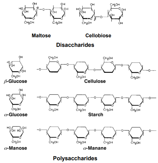
component of the cell walls of some microorganisms. It is a homopolymer
of N-acetyl-D-glucosamine. Both cellulose and chitin are insoluble in water.
Starch and glycogen are the basic reserve polysaccharides. Starch is
met in two forms — α-amylose and amylopectin.
Corn-bean plants, potatoes, etc. are especially rich in starch. Under the
action of enzymes starch decomposes to glucose, which is utilized in
metabolism. Glycogen is a reserve polysaccharide in the animal tissues and
organs. In larger quantities it is accumulated in the liver and muscles. It is easily
hydrolized by α- and β-amylase with decomposing to glucose and maltose.
Except with each other, carbohydrates form stable bonds with proteins,
nucleic acids, lipids, etc. Thus formed compounds, especially glycoproteins,
nucleosides and glycolipids, perform important biological functions.
All stated in Section 2. 7 gives only an idea about the structural
organization and importance of proteins, nucleic acids and carbohydrates
considered to be basic building blocks of the living systems. It does not
have for an object to exhaust them as problems from chemical and
biochemical point of view, since this is a huge subject, discussed in detail in
the special literature. Possibly, in the course of time some current views on
their role and importance will alter.
Noteworthy is the fact, that by forming the biopolymers —
polypeptides, polynucleotides and polysaccharides — the binding of single
monomers is accompanied by a removal of one water molecule (H₂O). The
mechanism is extremely similar (Fig. 2–52). A question arises: why these
processes are so similar and for what reason only H₂O is removed in them,
and whether it had been a matrix for their formation? Let hope that in near
future quantum physics and chemistry will answer this question, which will
throw light upon the general principles of organization of the living matter.

Page 124 Read More »

Page 123

Among disaccharides, saccharose, maltose and lactose are known
better. Saccharose, known also as cane-sugar, consists of two connected
monosaccharides — glucose and fructose. Maltose contains two residues
of D-glucose, while lactose contains D-glucose and D-galactose. Lactose is
found in milk and is called milk-sugar.
Raffinose is a representative of trisaccharides. Large quantity of it is
found in sugar beet. It consists of fructose, glucose and galactose residues.
Polysaccharides (also called glycans) are high-molecular compounds
with single or branched chains. They can be built of equal monosaccharide
units (homopolysaccharides). They are subdivided into structural and
reserve.

Figure 2–51. Binding of monosaccharides and formation of di- and 
polysaccharide chains.

Cellulose and chitin are the most spread structural polysaccharides.
Cellulose is widely spread in the plant kingdom. It participates in building
the wood, cotton fibres, cell walls of different lower and higher organisms. It
consists only of D-glucose residues. Chitin serves as a basic structural
element of the outer solid coat of most insects and crustacea, as well as a

Page 123 Read More »