book

Page 152

are obtained as a result of interspecies hydridization — allopolyploids.
Haploid genome mutations are obtained from the diploid or polyploid forms.
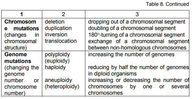
In aneuploid mutations the changes can affect one or several
chromosomes by increasing or reducing their number. Depending on that
they are: trisomia — increasing the genome by one chromosome (2n+1);
tetrasomia — increasing the genome by two chromosomes (2n+2);
monosomia — reducing the genome by one chromosome (2n-1);
nullisomia — reducing the genome by two chromosomes (2n-2), etc.

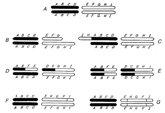
Figure 2–76. Chematic representation of the different types of 
chromosome mutations (After Rukmansky et al., 1984). 
A — two normal chromosome pairs; B — deletion; C — duplication; D — 
heterozygous translocation; E — homozygous translocation; F — 
heterozygous inversion; G — homozygous inversion

Classification according to phenotype. In accordance with this
classification, mutations are divided into morphological, physiological
and biochemical. Morphological mutations are connected mainly with
changes in the shape and colour. Physiological mutations cause changes
in the physiological processes running in the cells. As to the biochemical
mutations there belong the qualitative and quantitative changes in the
synthesis of certain substances or group of compounds. The latter are well
studied on microorganisms in auxotrophic mutations characterizing by
specific requirements for certain compounds necessary for their growth and
development.

Page 152 Read More »

Pge 151

Gene mutations. They are a result of changes in the primary structure
of DNA or RNA (when it is the basic genetic material) within the framework
of one gene. They are divided into point and block. Point mutations are due
to the changes of single nucleotides in the molecules of DNA or RNA. The
changes can be replacing of one nucleotide by another (transition),
dropping out of one nucleotides (deletion) and inserting of nucleotides
(insertion). Block mutations include a greater number of nucleotides or
whole segments of the polynucleotide chain of gene. They can arise
because of dropping out of certain segment (deletion), shifting of a segment
to other place in the gene (translocation), its turning to 180° (inversion) and
repetition of a nucleotide group in one gene (duplication).
Chromosome mutations (aberrations). This type of mutations arise
as a result of changes in the structure of chromosomes and can be
observed under light microscope. They are intrachromosomal and
interchromosomal. Intrachromosomal mutations are due to dropping out of
terminal or inner parts of the chromosome (deficiency and deletions),
doubling of chromosomal parts (duplications), turning to 180° of
chromosomal parts (inversions), and the interchromosomal — to exchange
of parts between two homologous chromosomes, which can be ordinary or
reciprocal (Fig. 2–76).
Genome mutations. They are a result of changes in the number of
chromosomes or in the genome as a whole. As it was already mentioned,
the haploid set of chromosomes (1n) is a fixed number. Except haploid, the
chromosomes set can be diploid (2n), triploid (3n), tetraploid (4n), etc.
Genome mutations are subdivided into three types: polyploidy or
euploidy — with increased genome number; haploidy — with reduced
genome number; aneuploidy (heteroploidy) — connected with changes in
the number of chromosomes. If increasing the genome number is within
one species, the obtained mutations are called autopolyploids, and when

Pge 151 Read More »

Page 150

useful economic features. The harmful consequences of mutagenic factors
are object of study by Ecological genetics.

Classification of the Mutations

It should be definitely said that up to now there is no comprehensive,
uniform and generally accepted classification. Those existed in the
literature can be reduced to three main groups: a) according to the way of
their arising; b) according to genotype; c) according to phenotype.
Classification according to the way of their arising. Depending on
that they are natural (spontaneous) and artificial (induced). Natural or
spontaneous mutations arise without human participation, under the influence
of different natural factors — earth and cosmic radiation, chemical
substances and other components of surroundings, changes of genetic
material, as well as some metabolites in the cells obtained as a result of
biochemical reactions. Artificial mutations arise through purposeful influences
by man using different physical, chemical and biological factors. That is why
they are called induced. On principle, there is no difference between
spontaneous and induced mutations, since their genotype and phenotype
appearance is similar. They differ only in mutation frequency, which in
spontaneous mutations is lower compared to that in the induced ones.
Classification according to genotype. It includes the changes in the
structure of genes and chromosomes, as well as in the genome, that lead
to changes in features and characteristics of the cells and organisms.
According to this classification mutations are divided into gene,
chromosome and genome (Table 8).

Page 150 Read More »

Page 149

gaining resistance against viruses, antibiotics or toxic substances, various
biochemical deviations, etc.

Mutation Theory

The observed sudden changes in morphology and hereditary features of
the organisms have attracted the attention of investigators. Some of them
have directed their attention to searching and elucidating the reasons for
their appearance. In 1899 the Russian botanist S. I. Korzhinsky has arrived
at the conclusion that these changes arise unevenly, without any transition
between the initial and newly form. The foundations of an orderly theory
has been laid by the Holland botanist Hugo de Vries (1901, 1903). For the
first time he introduced the term mutation to denote sudden and saltatory
hereditary changes.
Even now some of the basic principles of the mutation theory of H. de
Vries are valid. The most important of them are as follows: a) the mutation
arises suddenly, without any transitions; b) the newly forms are completely
constant, i.e. resistant; c) the mutations, unlike the non-hereditary changes
(fluctuations), are qualitative changes; d) mutations are realized in different
directions and can be useful, as well as harmful; e) same mutations can
arise over again.
W. Bateson (1902, 1909) have tried to elucidate the nature of
mutations by relating them to the hereditary factors of Mendel. Later on
Muller (1922) has arrived at the conclusion that mutations are closely
related to the nature of the gene. In 1925 Nadson and Filipov discovered
the mutagenic effect of radium rays on yeasts. Convincing evidence of the
effect of ionizing radiation on heredity and X-ray capability to cause
mutations are given by Muller (1927, 1928 a, b) in Drosophila and by
Stadler (1928 a, b) in barley and maize. There are laid the beginnings of
the experimental, i.e. induction mutagenesis. Also in the thirties, the
mutagenic effect of ultraviolet light is discovered.
Besides by ionizing radiations, mutagenic effect is manifested by some
chemical substances. Intensive research on them begins with the works of Ch.
Auerbach in England and I. A. Rapoport in USSR. The mighty mutagens
found, inducing more than 30—60% mutations, were called supermutagens
(see Rapoport, 1966).
Using the ionizing radiations and chemical mutagenic substances, as
well as their additive effect by combining, increased the power and the
possibilities of experimental mutagenesis. It began its considerable
progress as scientific branch. That not only encompassed theoretical
problems related to arising of mutations, their specificity and frequency, the
nature of gene and the character of chromosomal aberrations, but also
found practical application. There were obtained many new mutant forms in
different kinds of plants, animals and microorganisms, some of them with

Page 149 Read More »

Page 148

Mutation Changes – Basic Mechanism for Augmentation of the Gene Fund and Diversity of Living Organisms

Section 2.9. Variability is at the root of the very essence of nature. It is inherent in the
living, as well as in the non-living nature. Different are only the modes and
mechanisms of its realization. The changes in the living nature can be
temporary and persistent. Let pause on some of their characteristic features.
Temporary changes exist only during of the individual development of the
cells and organisms. After eliminating the reasons, caused their appearance,
they recover its initial form. Such changes are called modifications. They
have a temporary character and are not inherited.
Persistent changes are inherited. Because of that they are called
inheritable. They can arise as a result of genetic recombinations in case of
fusion of generative cells (fertilization), exchange of hereditary material
(transformation, transduction and conjugation), the influence of different
mutagenic factors, and others still unknown reasons. Sudden hereditary
changes are called mutations.

Prerequisites for the Generation of Idea of Mutation Changes

Sudden changes in the morphology and qualitative properties of naturally
existed plant and animal organisms are observed by people for ages. In
1590 the German pharmacist Sprenger from Heidelberg has noticed a
new form among the sown fields of the plant snake milk (Chelidonium
majus
) different from the other plants. Unlike the normal plants whose
leaves have had round lobes the new form has possessed cut leaves and
petals. He has called it Ch. laciniatum. In 1851, among usual strawberry
plants with trilobate leaves (Fragaria vesca), the French gardener A.
Duchesne has found a plant with unilobar leaves. He has called it F.
monophila.
The Chinese emperor Kang-Xi (1654—1722) has discovered
an early rice of better regarding grain qualities, become known as
“emperor’s”. Similar uneven changes are also observed in animals. In the
ranch of Ankon (Massachusetts, USA) a lamb with a long backbone and
short legs has appeared, originating Ankon breed of sheep (see
Rukmansky et al., 1984).
These is only a part of the curious cases of sudden hereditary changes
described in the literature. At that time it has been impossible to find a
scientific explanation. Their number is great and continues to increase.
Everybody knows that it is possible in a flock of white sheep a black lamb to
be born, in a flight of black swallows to meet one white for “luck”, in a
collection of Drosophila with red eyes to observe an appearance of one
with white (the case in the Laboratory of T. Morgan), a transformation of
antennae in some insects into legs, a change of the morphology in bacterial
colonies and loss or acquisiton of virulence by some cells in them, or

Page 148 Read More »

Page 147

There are also other cellular components — spherosomes, episomes,
lysosomes, peroxysomes, vacuoles, etc. Some authors consider them
organelles, others — organoids. A generalized idea of plant eukaryotic cell
is given in Figure 2–75.

image

Figure 2–75. Thin section of a cell from the root tip of wheat plant (After 
Brian Gunning; From Alberts et al., 1986). 
1 — cell wall; 2 — chloroplast; 3 — Golgi apparatus; 4 — 
mitochondrion; 5 — vacuole; 6 — nucleus; 7 — nucleus; 8 — 
ribosomes; 9 — endoplasmic reticulum. 

From all stated in this Section it is not difficult to arrive at the logical
conclusion that studying the living matter (objects) should be realized by
means of methods adequate to it, i.e. in vivo. It is quite possible that
cytoskeleton does not exist really, and its part is played by the cytoplasm in
a liquid phase which in its essence bear a resemblance to a membrane-encircled
drop of “primary bouillon”, where the individual cellular organelles
are freely located and they function being connected in a common system.
There is a real necessity of introducing new methods in order to achieve in
vivo
tridimensional picture of intracellular structures (like the holography).
This is the only way to avoid the non-conformity with the reality, and the
investigators will have a real idea of the building and functioning of cells.

Let us hope that during coming XXI century this dream will become usual
practice.

Page 147 Read More »

Page 146

Cytoskeleton

The term cytoskeleton directs the attention to the presence of some
locomotory system in the protoplasm, which has to determine the shape
and motion of cells. The high resolution of electron microscopy made it
possible to get some idea about it (Fig. 2–74 A, B).

image

image

Figure 2–74. Electron micrographs of cytoskeleton. 
A — cytoskeleton in fibroplasts, obtained after preliminary extracting the 
non-ionic detergents. The greater part of straight threads forming loose 
fascicles orientated from left to right are active filaments. The dense net 
in the middle of preparation is formed chiefly by intermediate filaments 
(Courtesy of Heuser and Kirschner, 1980) 
B — cytoskeleton in axon of rat, without preliminary extracting the 
detergents (Courtesy of S. Tsukita and H. Ishikawa; From Alberts et al., 
1986)

It is still unclear whether this fine net of filaments (threads), forming the
cytoskeleton, really exists in the cytoplasm of living cells or it is a result of
aggregation of soluble macromolecules during the process of fixation and
dehydration of preparations. That directs to the idea about the failure of using
such methods of investigation and the possibly great differences between
fixed and in vivo studies of the cells. There is a deep sense in the jocular
answer to the question: what is a biologist? According to the authors of the
joke — biologist is a person, who kills the living thing to study it!

Page 146 Read More »

Page 145

image

Figure 2–73. Polyribosomes from rabbit reticulocytes (Courtesy of 
A. Rich; From Levine, 1968). 

The biochemical composition of ribosomes includes mainly RNA and
proteins with very small quantities of lipids. In its essence they are
ribonucleoproteins — 60% RNA and 40% proteins, which are similar to the
nuclear histones. The proportion RNA/protein is of the same order for both
subparticles. The rRNA contained in them differ in sedimentation coefficient
and molecular weight. The small subparticle contains rRNA from 16S to
18S with molecular weight of 0.55—0.70×10⁶ daltons, and the large one —
from 23S to 28S with molecular weight of 1.2—1.7×10⁶ daltons varying in
different species. In the large subparticle one more kind of rRNA was
discovered — 5S, with a molecular weight of about 3.0×10⁴ daltons.
Still, quite a lot are the non-elucidated questions about the structural
organization of ribosomes, the presence and functions of different rRNAs in
them, etc. What is considered undoubted is, that they are the “cell plants”
for synthesizing proteins. Their key position in this process is the reason for
the great interest in them and subsequent numerous investigations.
Especially important is the question on the genesis of ribosomes in the
process of evolving the living systems and about the way of protein
synthesis in precellular forms before their appearance. There are two
possibilities: the first — they are a result of intracellular differentiation; the
second one — they have been included from outside, i.e. earlier they have
existed independently. Clarifying this question will shed light on some
aspects of the problem about the origin and evolution of life.

Page 145 Read More »

Page 144

image

Figure 2–72. Ribosomes in a bacterial cell. x 300 000 (After Oparin, 1966).

As structural units containing RNA and proteins, for the first time they
have been separated in homogenized cytoplasm of animal cells by Claude
(1943) and called microsomes. Under electron microscope they are
observed by Palade (1955). As early as next year the presence of a great
quantity of RNA was proven in them (Palade, Siekevitz, 1956 a, b).
By using the methods of gradient centrifugation and labelled atoms
many questions about ribosome structure and functions were clarified. It was
established that the ribosomes of prokaryotic cells (diameter 15—20 nm)
have a sedimentation coefficient about 70S and molecular weight of 2.5—
3.0×10⁶ daltons, and these of eukaryotic cells (diameter 20—30 nm) have a
sedimentation coefficient about 80S and molecular weight of 4—5×10⁶
daltons. The two types of ribosomes (prokaryotic and eukaryotic) have a
groove dividing them into two different subparticles — small and large. For
example 70S ribosomes consist of 30S and 50S subparticles, and 80S — of
40S and 60S. In a presence of Mg again they can form the initial 70S and
80S structures.
Sometimes ribosomes are observed in the form of small groups or
spiral structures, which have been called polyribosomes or polysomes
(Fig. 2–73). It is considered that in this way the effectiveness of using
mRNA is increased, since on them several polypeptide chains can be
synthesized at the same time. But, each one of them is an independently
functioning unit. The building of ribosomal subparticles is realized by a self-assembly
of RNA and protein molecules, at that corresponding rRNA
serves as skeleton of their structure.

Page 144 Read More »

Page 143

image

Figure 2–70. Cheme, elucidating the common features and differences 
in mitochondria and chloroplast structures.

image

Figure 2–71. Chloroplast of Chrysanthemum segetum in course of 
division (After Lance, 1958; From Kirk and Tilney-Bassett, 1967).

Ribosomes

They are small spheric formations (Fig. 2–72), extremely uniform in shape,
structure and chemical composition. Their dimensions vary from 15 to 35 nm.
They are observed in all kinds of cells — prokaryotic and eukaryotic. The
ribosomes are freely located in cytoplasm or attached to the outer
membranes of some organelles — mitochondria, chloroplasts, endoplasmatic
reticulum, etc.

Page 143 Read More »