book

Page 82

“special” chromosome is present in the cells of the male individuals which
was called Y-chromosome. Since it proved that these two types of
chromosomes are connected with the determination of sex they were called
sexual chromosomes.
Wilson and Stevens have suggested a comparatively simple mechanism
of distribution of the sexual chromosomes in the process of cell division. They
have assumed that each egg and the half of the spermatozoa have an X-chromosome.
The other half the spermatozoa contains the Y- chromosome.
When the egg is fertilized by a spermatozoon with an X-chromosome a
zygote of two X-chromosomes (XX) is obtained which gives rise to an
individual of the female sex. When the fertilization is carried out by a
spermatozoon with a Y-chromosome then the zygote has the two types (XY)
and an individual of the male sex is developed from it (Fig. 2–14).

image

Figure 2–14. Scheme for determining of the sex depending on the 
presence and combination of the X- and Y-chromosomes in the 
migratory short-tentacled locust (Dichroplus silveira guidoi) with four 
chromosome pairs (2n=8). (After De Robertis et al., 1973). 

Page 82 Read More »

Page 81

Chromosome Theory of Heredity and Organization of the Genome

Section 2.6. The chromosome theory of heredity is one of the great
achievements in biology. The rules of inheriting the features in the next
generations discovered by Mendel have laid the ground for its buildup and
further development. In his time these discoveries have not found their due
explanation since cell division and meiosis responsible for the formation of
the gametes (i.e. the sexual cells) taking part in the fertilization have not
been known yet (see Chapter 3, Section 3. 2 and 3. 4).
In 1884 K. Nägeli has admitted hypothetically the existence of
idioplasm as a specific hereditary substance. Almost simultaneously E.
van Beneden, E. Strasburger, O. Hertwig, R. Köllicker, A. Weissmann,
etc. have come to the conclusion that Nägeli’s idioplasm could be found
in the chromosomes.
The chromosome-heredity link has greatly captured the imagination of
many researchers. The cytological studies on the behaviour of the
chromosomes, in the process of cell division and meiosis have shown that
the sexual cells — spermatozoa and eggs — possess a single (haploid) set
of chromosomes and the fertilized ones — the zygotes, contain a double
(diploid) set of chromosomes. T. Boveri and W. Sutton have related these
results to the rules observed by Mendel in the inheritance of the features in
the process of hybridization in peas.
In his classical paper “The Chromosomes in Heredity” the American
biologist W. Sutton (1903) has for the first time paid attention to the fact that
the diploid chromosome set consist of two morphologically similar haploid
sets of chromosomes and in the process of meiosis the gametes receive
one haploid chromosome from each homologous pair. He has made the
assumption that the genes are a part of the chromosomes, and being
based on the data available at that time from the cytological studies has
explained some of Mendel’s results in the hybridization. This work has
played a great role in the creation of the chromosome theory and in
connecting the cytology with genetics.
The view that heredity is to be found in the chromosomes and the
chromosome number is specific and exactly determined for each separate
species of organisms has been already shared by most of the researchers.
Around 1890, however, a chromosome has been discovered which was not
always observed in two copies in the cells. This chromosome was
interpreted as an additional or sexual chromosome (McClung, 1901) and
has later become known as X-chromosome. Its significance has been
clarified by E. Wilson and his student Stevens. They have assumed that
there are normally two X-chromosomes in the cells of the female individuals
and in the cells of the male individuals — only one. Besides, it has been
established that in some species (the human including) yet another

Page 81 Read More »

Page 80

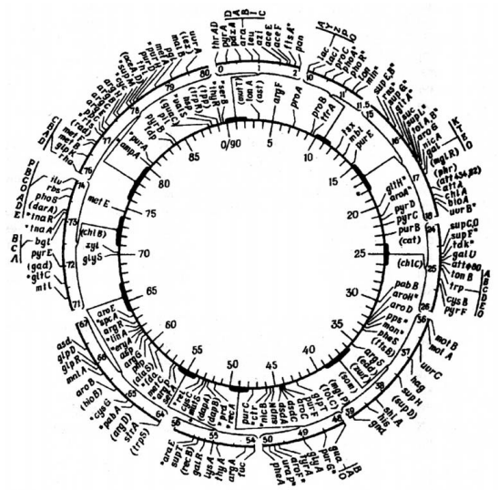
It is very difficult practically (and may be unnecessary) to survey and
discuss the results from the numerous studies on the transfer of genetic
material from the donor cell into the recipient cell, the various
recombinations resulting from that act and the mechanisms controlling it,
the exchange and binding of the genetic markers, competence or
incompetence of the recipient cells, phage conversion, the kinetics of these
processes, etc. All these problems are considered in detail in the
specialized literature. It is however necessary to note that the discovery of
transformation, conjugation and transduction has served as a solid proof for
the assumption that hereditary information is confined to the DNA molecule.
This has played an essential role in the building of the chromosome theory.
In the period 1944—1950 comparative measurements of the DNA
content in the nuclei of cells from different organisms were made. The data
have shown that the quantity of DNA in a complete chromosome set
remains unaltered for all cells of the given organisms while the protein
content is subject to significant changes. On the basis of these results
some authors (Boivin et al., 1948; Vendrely, Vendrely, 1948, 1949; Swift,
1950 a, b, etc.) have forwarded the hypothesis for the permanent amount of
DNA which in polyploid cells is increased multiply to ploidity (2n, 3n, 4n,
etc.) and in sexual cells is reduced to half the diploid chromosome number.
At present, data is available about the intraspecific differences in the DNA
content which are not to be discussed.
At the same time the American biochemist E. Chargaff (1950) has
established that the sum of the purine bases (adenine, guanine) is equal to
the pyrimidine bases (thymine, cytosine). Besides he has discovered that
the quantity of thymine (T) is equal to that of adenine (A) and the one of
guanine (G) is equal to that of cytosine (C), and these ratios varying in the
different types of organisms. This fact has served as a ground for the
assumption that the differences among DNA molecules are greater than it
has been admitted in the already mentioned hypothesis for the
tetranucleotide structure of nucleic acids. These data known as the
Chargaff’s rules have been implemented in the disclosure of the spatial
structure of DNA.
In 1953 Watson and Crick (Watson, Crick 1953 a, b) have published
their model of the DNA double helix and thus have shown the possibilities for
its self-replication which has marked the onset of modern molecular biology
and genetics. This problem will be considered in more detail in Section 2. 7
(Nucleic acids).

Page 80 Read More »

Page 79

name of transduction. The next year Zinder and Lederberg (1952) have
established that the agent causing the transduction is indiscernible from the
particles of the moderate bacteriophage PLT-22. At the same time Hershey
and Chase (1952), by means of labelled atoms (³⁵S and ³²P) and using the
bacteriophage T2 as the object of study, have proven that only the DNA
molecule penetrates into the bacterial cells and its protein envelope is left
behind.
In a few years time Fraenkel-Conrat and co-authors (Fraenkel-Conrat,
Williams, 1955; Fraenkel-Conrat, 1956; Fraenkel-Conrat, Singer, 1957 a;
Fraenkel-Conrat, Singer, Williams, 1957 b) have established that not only
DNA but viral RNA can also play the role of genetic material. The studies of
Gierer and Schramm (1956, 1957) are directed into that trend. It has later
become clear that the viral DNA and RNA molecules can be both single
stranded and double stranded.
At that time a lot of researchers have made effort to prove that DNA
alone is the component responsible for transduction and conjugation. This
problem is very essential in biology and it therefore will not be left
uncommented.
In his book “The Genetics of Bacteria and their Viruses” W. Hayes
(1964) pointed out that “it turns out that about 3 per cent of the total phage
protein is infected along with the DNA. This protein, however is not part of
the phage envelope but comprises those polyamines and other constituents
which are contained within the phage head and are liberared by osmotic
chock (internal proteins)”.
With experiments in which transforming preparation with a more
thorough purification of DNA have been used, it has been shown that
transforming activity has increased and progressive release of proteins,
other serologically active substances and RNA has not reduced its potency
at all. According to Hayes “The transforming principe, if not DNA, must
therefore be a substance so bizarre that its nature is difficult to imagine””
Here, most properly the question arises: what are these
physicochemical forces and biological mechanisms which roughly for a
minute’s time (the time needed for the passing of the DNA molecule
through the channel thus formed into the recipient cell) could “purify”
thoroughly the DNA molecule from proteins and all sorts of other
components so, that it could in an absolutely pure state be incorporated
into the genome of the bacterial cell and quickly (for about 12 min) to
reproduce and yield the first numerous infections agents.
In my opinion the transforming factor would not have been of so
“bizarre” a nature if it was assumed that together with the DNA also other
components were passing into the recipient cell, which is natural for such a
biological process. The answer to this question is not only necessary but it
is obligatory as well, so that the doubts that this is possible be cleared up.

Page 79 Read More »

Page 78

stopping the conjugation at the appointed time (intervals) developed by
Wollman and Jacob provided the possibility for mapping the genes and
constructing of genetic map of E. coli was made, whose circilar DNA
molecule is divided into 90 equal segments (minutes) — Figure 2–13. Such
maps have been made also for other biological objects.

Figure 2–13. Genetic map of Escherichia coli made upon scale. The
map is divided into 90 intervals of one minute each. The symbols and
the description of the different genetic markers are given in Appendix 1
(After Taylor and Thoman, 1964; Taylor, 1970).

In 1951 Lederberg and co-authors have discovered in Salmonella
typhimurium
a transfer of genetic material by viruses leading to
recombination that have been earlier observed in E. coli K-12 (Lederberg,
Tatum, 1946 a, b; Tatum, Lederberg, 1947). This process was given the

Page 78 Read More »

Page 77

The contact between the F⁺ and F⁻ cells is accomplished by F-pilli. If the
F-factor is integrated in the bacterial chromosome (it can also be found in a
separate structure as is the case with the plasmids) and has a high
frequency of recombination, then the strains are designated with the
symbols Hfr (high frequency of recombination).

image a

image b

Figure 2–12. Electron micrographs of conjugation in cells of Escherichia
coli (After Anderson et al., 1957; Wollman and Jacob, 1959).

(A) Conjugation between K-12 Hfr H anf GF⁻. The K-12 Hfr bacterium is
elongated and in the process of division, while the G F⁻ bacterium is
thick and rounded. The two bacteria are connected by a cytoplasmic
bridge. In the contact area the cell walls seem obliterated. The K-12 Hfr
bacterium is flagellated and is therefore mobile, while the G F⁻
bacterium has no flagella and is not mobile; (B) Conjugation between
bacteria Hfr F⁻λʳ and F⁻λˢ. The two bacteria are close to each other. In
the contact area the walls seem obliterated. The F⁻ bacterium is in a
process of division and is covered by a number of bacteriophages λ,
which are fixed by the help of their tails.
The transfer of genetic factors (markers) takes place under certain
conditions and for a definite period of time. The method of starting and

Page 77 Read More »

Page 76

Griffith has carried out his experiments on mice and guinea pigs that
have been infected with pneumococci (Streptococcus pneumoniae). One of
the strains was strongly virulent causing serious disease of the respiratory
organs, while the other one was avirulent and thus harmless. The virulent
strain cultivated on an agar nutrient medium has been forming typical
brilliant colonies with a smooth surface. The avirulent strain under the same
conditions has been forming turbid colonies with a roughly surface. Except
for this, the two strains were differing in yet another genetically determined
feature — the virulent one had a polysaccharide capsule, while the
avirulent did not form such a capsule.
When the experimental animals were injected with the avirulent strain
they have all remained alive, while when the pneumococci introduced in
them have belonged to the virulent strain the animals were all dead. The
variation in which mixed injections were given containing the native
avirulent strain and killed by heating pneumococci of the virulent one
proved to be more intriguing from a biological point of view. Most
unexpectedly all animals died. The pneumococci isolated from them have
been forming colonies typical of the virulent strain and displaying
polysaccharide capsules. Griffith has suggested that this transforming
factor could have been the polysaccharide itself.
The reason for this phenomenon was later elucidated by the American
microbiologist O. T. Avery and co-workers (1944). It has been established
that DNA extracted from capsulated smooth colonies (S-type) has
transformed the uncapsulated bacterial cells of the R-type (rough) into
capsulated ones with well-expressed virulent capacities. This phenomenon
was given the name of bacterial transformation. A great number of other
researchers have shown that it is characteristic of lots of bacteria.
After two years J. Lederberg and E. Tatum (1946 a, b) discovered a
sexual process in bacterial cells leading to genetic recombinations. This
process was called conjugation. It was studied in detail on Escherishia
coli but was confirmed also on other bacterial strains, which is a proof for
its wide spreading in prokaryotic cells. It is quite possible for this
phenomenon to be a major biological tool for exchange of genetic
information in the independently existing unicellular organisms.
As a process conjugation has become the target of many studies
(Nelson, 1951; Hayes 1952—57; Clowes, Rowley, 1954; Wollman, Jacob,
1955—58 a, b; Jacob, Wollman, 1956, 1958, etc.). The monographs of E.
Wollman and F. Jacob (1959, 1961) are remarkable in that respect. By the
help of electron microscopy the existence of cytoplasmic bridges in
bacterial cells and the exchange of genetic information between them has
been convincingly proved (Fig. 2–12 A, B).
The studies in that direction has disclosed the existence of “sexual”
differences in bacterial cells called F-factors. The cells which donate
chromosomal markers are designated F+
and the cells receiving them — F⁻

Page 76 Read More »

Page 75

they are not to be discussed here, being out of the subject of this book.
Till the middle of the XX century the studies on nucleic acids have been
episodic and in small numbers. The attention was mainly directed to the
discovery of the structure and biological importance of proteins. It was already
known that protein molecules represent long amino-acid polymer chains that
are very variable in shape and size and are of high-molecular weight. Because
of these important biochemical characteristics of theirs, a kind of genetic
control over them by some proteins (especially the enzymes) was admitted.
This has led to the assumption that if not enzymes were the genes
themselves, then their structure could have been determined by proteins.
At this time nucleic acids were considered short and simply structured
polymer chains that could not be capable of preserving and transmitting
hereditary information. The majority of authors were supporters of the
tetranucleotide hypothesis, which has postulated that the four types of
nucleotides (including the nitrogenous bases — adenine, thymine, cytosine
and guanine) are present in the DNAmolecule in equimolar quantities of
identical recurring monomers, similar to these in glycogen. That is why it was
thought that DNA does not have the capacity to ensure the gene variety
needed for determination of the thousands of hereditary features displayed by
living organisms. The noted English crystallographer W. T. Astbury has stated
“that the symmetrically situated nucleotides in DNA are only outlining the
framework in which the amino acids of the protein genes would extend into
chains before they reproduce themselves”.
The formation of the concept of proteins as bearers of genetic
information resulted from an enormous number of studies carried out in that
period. Especially important are the X-ray diffraction analyses of proteins and
enzymes (hemoglobin, myoglobin, pepsin, myosin, etc.) made by
researchers such as J. Bernal, L. Pauling, M. Perutz, J. Kendrew, etc. In
his famous book “The Nature of the Chemical Bond” Pauling (1960) has
offered a summary of his studies on the structure of proteins and the αspiral model,
which will be discussed later in Section 2. 7 (Proteins).
An interesting biological phenomenon was discovered by the
English bacteriologist F. Griffith (1928). His experiment is a classic in the
history of genetics and a turning point in proving that DNA represents the
genetic material and therefore it will be spelled out in detail (Fig. 2–11).

Figure 2–11. Schematic representation 
of the Griffith’s experiments with 
pneumococci.

Page 75 Read More »

Page 74

the genetic continuity of chromosomes do not depend on the degree of
quantitative changes of chromatin”.
The “mysterious” substance incorporated in the chromosomes has
become a subject of special attention. Around 1890 R. Altmann and A. Kossel
have proven that nuclein consists of two components — protein and an acid
rich in phosphorus which has later been known as nucleic. Let remember,
Miescher has suggested that the substance isolated by him is an acid in its
nature. He has established the phosphorus content in this substance to 9.5
per cent. It has taken a long time to understand that there are two kinds of
nucleic acids. The one isolated from the thymus of calves and the other —
from yeasts. This has created the incorrect idea in some authors that the one
is an animal acid and the other — a plant one.
By chemical analyses in 1900—1903 the research groups of A. Kossel
in Germany and P. A. Levene in the USA have clarified this
misunderstanding. Kossel has established that the thymic nucleic acid contains
adenine, cytosine, guanine and thymine while the yeast one contains uracil
instead of thymine. The presence of uracil in the yeast nucleic acid has
been established by A. Ascoli before him. Levene on his part has
established that the carbohydrate component of the yeast derived nucleic
acid is ribose and the one of the thymus — deoxyribose (see Fig. 2–48),
which has not been known up till then. The ribose containing nucleic acid
was given the name ribonucleic acid (RNA) and the deoxyribose
containing one — deoxyribonucleic acid (DNA).
A major contribution to the cytological visualization of DNA was made
by the German chemist R. Feulgen. In 1924 Feulgen and Rossenbeck have
proposed a histochemical method for a selective visualization of DNA in the
nuclei of cells of animal and plant origin. It was later applied to
microorganisms. This method became known as the Feulgen reaction. It
consists of two stages: a) hydrolysis with acids (mainly HCl) as a result
from which the bonds of the nitrogenous bases of DNA are broken and
aldehydes are formed; b) placement in Schiff’s reagent (fuchsine
sulphurous acid), dehydration in ethanol and xylene, and the presence of
DNA is recorded by the red-violet structures formed in the nucleus.
In spite of the numerous studies on the Feulgen reaction its specificity is
not clear to the present day. There is no convincing answer as to why
aldehyde groups are formed from DNA and not from RNA. The authors have
expressed an opinion that in hydrolysis the bonds between the purine bases
and the carbohydrate components of DNA are broken more easily than in
RNA. Some researchers, however, have shown the release of great
quantities of RNA in acid hydrolysis. Besides, in some samples the reaction
is positive, and in others it is negative i.e. there is no staining. The negative
answer cannot be accepted as a proof for the absence of DNA in the nuclei.
There are also other methods for cytological studies, which are based on
other reagents for fixing and staining. They are being successfully applied but

Page 74 Read More »

Page 73

A. Weissmann and other authors. According to his opinion “the clue to the
solution of fertilization was lying in stereochemistry”.
The biological role of nuclein has begun to become clearer due to the
studies of a number of biologists and cytologists — O. Hertwig, W. Flemming,
E. van Beneden, etc. They have observed the penetration of spermatozoa in
eggs of various animal species (mainly sea urchin and sea star) after which
their nuclei have fused. A great interest was provoked by the thread-like
formations in the nuclei later called chromosomes (Greek: chroma — colour
and soma — body). Yet at that time Hertwig (1875) has arrived at the
conclusion that the chromosome number is doubled in the fertilization, and
Flemming (1880) has surmised that after the division of the chromosomes
along their axis each half goes to one of the daughter cells. In two years time
Flemming (1882) has described a division of epithelial cells from salamander
in which the chromosomes are clearly outlined. He has suggested the term
mitosis and has reached to the conclusion about the constant chromosome
number in the cells of a given species. Van Beneden (1883 b) has described
fertilization and division of cells in the parasitic round worms Ascaris. This
subject of study has proved very convenient since by contrast to other ones, it
has only two chromosomes in the nuclei of sexual cells which allows for the
easy observation under the light microscope. In this work of his van Beneden
has for the first time described meiosis — the reduction of the chromosome
number by half as opposed to the process of fertilization. Up till then the role of
the nucleus has remained unclear although it was already discovered. This
essential problem connected to the discovery of cell division will be later
treated again in Chapter 3 (Section 3. 2).
Nuclein has been actively occupying researchers minds. Cytological
studies in vitro as well as after fixation and staining of the cells have shown
a zone of a dense matter in the nucleus called chromatin. It was later that
the notion has arisen that in the process of division the maximally
condensed chromatin is shaped as chromosomes. At that time the giant
(polythene) chromosomes have been discovered in the salivary glands of
Chironomus (Balbiani, 1881), as well as the lampbrush type chromosomes
in oocytes of Siredon (Flemming, 1882). The idea has cropped up that
nuclein was the hereditary material in the observed chromosomes. In 1895
the American cytologist E. B. Wilson has assumed that nuclein is very
similar if not identical to the substance called chromatin. It has proven
however, that the quantity of chromatin varies with the timing of the cell
division cycle and the physiological state of the cells. In 1909 the German
botanist E. Strasburger has expressed his opinion that chromatin cannot
serve as hereditary material, since its content is considerably changing in
the process of cell development. This view of Strasburger was adopted by
Wilson and in the third edition of his book “The Cell in Development and
Heredity” (Wilson, 1925) he has stated that “individuality of organisms and

Page 73 Read More »