juliana

Page 172

image

image

image

image

Figure 2–88. Electron micrographs of some well-known viruses and 
phages (After Fraenkel-Konrat. 1985) 
1 — tobacco mosaic virus (18 x 300 nm); 2 — potato virus X (13 x 600 
nm); 3 — turnip yellow mosaic virus (diameter 29 nm); 4 — human 
herpes virus (about 200 nm diameter). The tubular capsomeres, 162 in 
all, are clearly distinguishable; 5 — adenovirus (diameter 80 nm) largely 
composed of hexones, with fibres (27 nm in length) attached to the 
pentons; 6 — phage φx 174 (diameter 27 nm, with spikes at the vertices 
of the icosahedron); 7 — phage λ (diameter of the head — 54 nm, tail — 

15 x 150 nm); 8 — phage T4 (head — 80—95 nm and tail — 16 x 110 
nm) with clearly visible neck, tail fibers, base plate, and spikes.

Page 172 Read More »

Page 171

of the phages into bacterial cells begins with their attachment through the
tails (see Fig. 2–12 B).
Great is the diversity of viruses in the living nature. In the special
literature on virology it is called Vira Kingdom with more than 3000 species.
It is imposible to be described and depicted adequately in a short exposition.
For that reason, only some represetatives will be shown (Fig. 2–88).

image

image

image

image

Page 171 Read More »

Page 170

1946; Newcobmbe, 1949; Lederberg, Lederberg, 1952). One more avalanche
of investigations occurred in this field. A new branch of biology appeared —
Virology.
The viruses can infect the cells of all kinds of plants, animals and
microorganisms. Their typical feature is that unlike the parasitizing
rickettsiae and bacteria they do not possess their own metabolite system.
That is why, for their developing and reproducing they need the biological
apparatus of recipient cell. Developing in the cells they destroy or lyse them
(Fig. 2–87). As a result the “hosts” perish.
Virulent phages immediately lyse the infected cells. They have only
one way of development, so-called lytic cycle. Also there exist moderate
phages (P1, P2, P22, λ, etc.). They can penetrate into recipient cell without
immediate lysing it and are transferred to its following generations. Such
bacteria are called lysogenic, and the phage — prophage. At a given
moment a prophage can change into virulent and proceed to lysis. The
reasons for that are unclear.

image

Figure 2–87. Electron micrograph of lysed culture of E. coli infected with 
T2 phage (After Stent, 1974).

Adsorbing the phages is closely connected with the cellular envelope.
Except by living cells. they can also be adsorbed by empty cellular
envelopes. Cells with destroyed envelopes (protoplasts) do not absorb
them, but they develop there if have been included previously. Penetration

Page 170 Read More »

Page 169

image

Figure 2–85. Localization of plant viruses (After Fraenkel-Konrat, 1974). 
Infectious loci appear 6 days after inoculating with tobacco mosaic virus 
(TMV). One half of the leaf surface is infected with a normal TMV (the 
larger spots), and the other half — with TMV-mutant (the smaller spots). 
Figure 2–86. Cycle of phage 
development (After Frey-Wyssling and
Mühlethaler, 1965).

The discovery of electron microscope in the end of third
decade of XX century made it possible to observe the viruses
(phages) visually. The first electron micrograph of a phage is made by
Ernst Ruska in 1940, and two years later — by S. Luria and T.
Anderson of T-even phages (T2 T4 and T6), parasitizing on E. coli.
Regarding the dimensions the phages were found to be very
small (about 0.1 nm, with a weight of 4×10⁻¹⁶ g), which is approximately one
thousandth compared to the bacterial host-cells. Their
development cycle is given in Figure 2–86.
The interest in viruses (particularly in phages) greatly increases.
especially after elaborating new methods of experimental analysis
and recording the recombinations and mutations in bacteria and
viruses (Luria, Delbrück, 1943; Luria, 1945; Delbrück, Bailey, 1946; Hershey,

Page 169 Read More »

Page 168

Are the Viruses Cells?

Section 2.11. Posing this question is not an act of curiosity. It is an essential
question. The viruses really exist. They contain DNA or RNA
and proteins (in some of them are established lipids, carbohydrates and
other components), cause genetic recombinations and mutations as it is in
the normal cells, reproduce and create numerous generations, but up to
now they still have not found a place in the evolutionary tree of living
organisms (see Fig. 1–2).
The existence of viruses has been predicted long time before to be
really observed. For the doctors in ancient Rome “virus” has meant poison
of animal origin, and the diseases caused by such poisons has been called
“viral”. Also the monstrous disease hydrophobia has been assigned to
them. In the Renaissance it has become clear that one should make
difference between non-contagious diseases caused by poisons and
contagious diseases caused by infectious agents. The term “viral disease”
has remained only for the contagious ones.
In XIX century as a result of the advance in microbiology it has been
established that infectious agents are bacteria, which really cause many
contagious diseases. Because of that, they have been incorrectly
considered “morbid viruses”. At that time bacteriological methods have not
enabled to observe and identify real viruses, in order to differentiate from
bacteria. L. Pasteur has known that hydrophobia in man is caused by a
specific living agent and it is transferred by animals (for example through
biting by a dog), but he has not succeeded to cultivate and isolate it on any
bacterial medium.
At the end of the century it has been found out that these infectious
agents are smaller than bacteria, since they have passed through the
filters which normally have stopped the bacteria known at that time. For
that reason they have been called “filtering viruses”.
The beginning of the intensive study of viruses starts with revealing
the cause of the mosaic disease in tobacco (Nicotiana tabacum). In 1892
D. I. Ivanovsky has established that its agent is invisible under
microscope, passes through porcellanous filters and does not grow on
usual nutrient media. In 1915 F. Twort has discovered that the viruses
infect not only the eukaryotic cells of higher organisms, but also bacteria.
Two years later (1917) F. d’Hérelle who has arrived to analogical
conclusion called them “bacteriophages” (from bacteria and phagos —
eat. swallow). For brevity they are called phages, W. Stanley (1935) has
succeeded in obtaining the tobacco mosaic virus (TMV) in crystalline
form. The localization of TMV is given in Figure 2–85. The term “virus”
as “contagium vivum fluidum” is suggested by Beijerinck (1898).

Page 168 Read More »

Page 167

such a case the admissible genetic autonomy of flagella is 20%, and the
dependence on the cell nucleus is 80%.
The presence in the cells of organelles having its own DNA, specific
RNA and ribosomes, and their capability to divide independently posed a
difficult dilemma before the investigators: eukaryotic cell — is it
elementary structure or endosymbiotic system?

This dilemma supposes two possible ways of evolutionary developing
of the eukaryotic cells: 1) through intracellular differentiation, i.e.
compartmentalization of the above-mentioned autonomous organelles; 2)
through endosymbiosis of protists existed independently earlier. The first
concept is supported by the successive theory, and the second — by the
endosymbiotic. Also, there exists so-called combination theory. According
to it eucists have as endosymbiotic, as well as successive origin. This
theory cannot answer the above dilemma, since accepting endosymbiosis
though to a certain extent, in reality it supports the endosymbiotic theory.
What is important in this case is whether the extranuclear DNA is a product
of the cell itself, or it is included in it by ancient ancestor.
Vast theoretical and experimental work was carried out to confirm one
or the other of the two basic theories. There were used different methods of
investigation — analyses of microfossils and DNA-containing organeles,
revealing similarities between their nucleotide sequences. making
analogies with contemporary independently existing protocists, studying the
genetic autonomy of individual organelles, etc. Most results obtained up to
now evidence in favour of endosymbiotic theory, but they still cannot prove
it completely.
Because of the importance of the problem, in 1980 and 1983 in
Tübingen (Germany) there were held 1st and 2nd International Symposia
on “Endosymbiosis and Cell Research”. As a result of these two forums a
new branch of biology appeared — Endocytobiology.
Having in mind contemporary rapid development of science and the
possibilities and methods of investigation in molecular biology and gene
engineering, one can expect creation of organelle cultures like the cellular
ones. Thus, there will be an answer to many questions related with the
extranuclear heredity. If the endosymbiotic theory is proven indisputably,
then according to W. Schwemmler (1977) “in the future it will play the same
role, as at its time the atomic theory for the chemistry”. Studying of the cell
will enter upon a new stage of its development. This on its part will impose
reconsidering the contemporary idea on the genome of eukaryotic cells in
the light of the obtained new data on their hereditary endowments.

Page 167 Read More »

Page 166

mitochondria it was established that organelle DNA-molecules are circular,
specific and differ from the average nucleotide composition of nuclear DNA.
That is used as a basis for their identification. It was proven that the
replication of DNA in the nucleus and that of chloroplasts are realized in
different phases of the cell cycle (Chiang, Sueoka, 1967). Also there
appeared reports about a presence of DNA in basal small bodies of the
flagella in some unicellular algae and Protozoa.
Extranuclear heredity which in many cases determines a non-
Mendelian type of inheritance, the presence of specific DNA, RNA and
ribosomes, as well as the differences during the replication of nuclear and
chloroplast DNA-molecules gave serious grounds to accept the existence
of genetic autonomy of some organelles participating in the composition of
eukaryotic cells. By means of modern methods numerous studies were
carried out in this field. As a result of them an opinion was confirmed, that
these organelles are endosymbionts originating from common protist
ancestors — bacteria and unicellular blue-green algae.
At present there exist the following ideas on the genetic autonomy of
some cellular organelles.
Chloroplasts. Circular DNA-molecule of the chloroplasts has a
molecular weight of about 1×10⁸ daltons, which corresponds to the
genome of lower bacteria like mycoplasms and rickettsiae. By means of
the available 250 000—300 000 base pairs the chloroplast genome may
code the synthesis of about 200 proteins with average molecular weight
of 40 000. In reality, according to the measurements, only 20% of the
genetic information is active. That means a synthesis of maximum 40—
50 proteins. If juxtapose the amount of their own proteins — about 50 to
100 — the corresponding minimum in protists determined by Kaplan
(1972), the chloroplasts code only 50% of their essential proteins. In this
case the proportion between chloroplast genetic autonomy and the
dependence on the nucleus is 50:50.
Mitochondria. These organelles also have circular DNA-molecules
with a molecular weight of about 1×10⁷ daltons, with dimensions less than
the average of now-existing protists. The mitochondrial genome is active
and with its 15 000—20 000 base pairs is capable to synthesize about 30—
35 proteins with average molecular weight of 20 000. If juxtapose the
mitochondria-own proteins with the total 100 proteins in protists, then
mitochondria code only 35% of their essential proteins, i.e. their genetic
autonomy is up to 35%, and the dependence on the cell nucleus is up to
65%.
Flagella. This is the third organelle in eukaryotic cells considered to
have its own DNA. It is accepted that DNA-molecule in the bases of
flagella (i.e. in the basal small bodies) having a molecular weight of about
1×10⁶ daltons, approximately equal to 10 000 base pairs, is capable to
code 20 proteins with average molecular weight of 20 000 daltons. In

Page 166 Read More »

Page 165

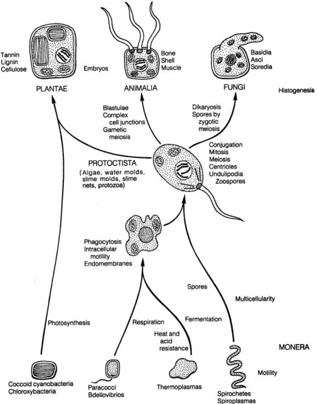
Figure 2–84. Model for the origin of eukaryotic cells by symbiosis (After 
Margulis, 1983). 

The decisive role in elucidating this problem belongs to the discovery
of genetic material (DNA) in some cellular organelles — chloroplasts (Ris,
Plaut, 1962; Chun et al., 1963; Sager, Ishida, 1963), mitochondria (Nass,
Nass, 1963 a, b; Luck, Reich, 1964; Schatz et al., 1964), etc. Mainly in

Page 165 Read More »

Page 164

Endosymbiotic Organization of Eukaryotic Cells

Section 2.10. It would be much easier for the investigators, if the existence
and evolutionary development of the cells had not been so long and
mutually dependent owing to contacts and interactions. These two
unavoidable circumstances have exerted their influence upon their
structural organization.
During last several decades the concept of endosymbiotic nature of
eukaryotic cells is gaining more and more adherents. The idea that some of
cell organelles are symbiotic inclusions is expressed by Schimper (1883)
more than a century ago. Mereschkovsky (1905), Famintzin (1907), Kozo
Polyanskii (1924) develop it further on the basis of their morphological
similarity with independently existed prokaryotic cells. As early as in 1868
Famintzin have arrived at the conviction, that chlorophyll grains resemble
Chlorella and Xanthella and tried to obtain pure cultures of chloroplasts.
Correns (1909) and Baur (1909) have described the first cases of
cytoplasmic heredity in Mirabilis jalapa and Pelargonium zonale. Ruth
Sager (1975) and Lynn Margulis (1983) are ardent adherents of this idea
with important contributions to its further development. The process of the
origin of eukaryotic cells by symbiosis is shown in Fig. 2–84.
Accumulating more data on cytoplasmic heredity made the
investigators try to find the reasons for this phenomenon. The Mendel’s rule
about a symmetrical segregation of hereditary features in gametes in
proportion 2:2 has been seriously violated in cases when the heredity is
transferred only in the female line asymmetrically, and the proportion of
gametes is 4:0. Besides, it has been established the presence of
autonomously replicated cytoplasmic structures (plasmids, bacteriophages,
etc.) moving from one cell to another being in close interrelations with the
chromosomes in nuclei, without any synchron with the phases of mitotic
processes realized in them. It had been impossible to explain these cases
from the viewpoint of chromosome theory, which in its classic aspect
defines the chromosomes in nucleus to be the only carriers of hereditary
features. There have been accepted two types of inheriting: Mendelian and
non-Mendelian.
It is necessary to emphasize that in the period 1910—1960 the data on
the existence of cytoplasmic genes have accumulated very slowly and very
often they have not been acknowledged. The literature on cytoplasmic
heredity has been considered a “spot” discrediting the science. Only in the
forties of our century, under the influence of a number of remarkable
discoveries in this field, it became possible to suppose a genetic autonomy
of organelles in the cytoplasm.

Page 164 Read More »

Page 163

The reparation trough excision is more complex. Here a complex of
enzymes participates and there is no necessity of daylight as a source of
energy. It is realized in dark. Its essence is an enzymatic recognition and
removal of the damaged part in one of DNA-chains, resynthesizing the
same under the action of DNA-polymerase and sewing up the rupture by
means of a specific group of enzymes called DNA-ligases. This mode of
raparation refers not only to pyrimidine dimers, but also to other kinds of
damages causes by UV-light, X-rays, γ-rays, alkylating agents, etc.
Postreplication restoring (reparation) is referred to the processes and
changes occurring during and after the replication of the damaged DNA.
That determines the importance of the connection of these processes
with S-period, i.e. the synthesis of DNA. It is supposed that some of the
enzymes, participating in the reparation processes through excision and
resynthesis of damaged DNA-segments, may play an important role in the
normal realization of genetic recombinations and in maintaining the
integrity of the two-chain molecule of DNA. Besides, it is accepted that
pyrimidine dimers do not block the replication of DNA but cause the
formation of gaps in the locuses of newly synthesized chains,
corresponding to the dimers in initial chains. Fujiwara (1972) has
established that the number of gaps corresponds to the number of
pyrimidine dimers. But the mechanism of their filling remains unclear.
All stated in this Section leads to the conclusion that the knowledge
about the mutation changes is insufficient. Most processes and
mechanisms of their realization are given schematically. That gives
grounds to consider this problem one of the “white spots” in biology.
Undoubtedly, the mutations are connected with the hereditary material,
the chromosomes and genome as a whole, respectively. However, the
chromosomes are built not only by DNA. As it was already mentioned,
also RNA and a large set of proteins participate in chromatin composition.
Before undoing this Gordian knot of biology, i.e. to elucidate the
connection and interactions among them objectively, the problem about
the mutation changes will be open.

Page 163 Read More »