image
Figure 2–85. Localization of plant viruses (After Fraenkel-Konrat, 1974).
Infectious loci appear 6 days after inoculating with tobacco mosaic virus
(TMV). One half of the leaf surface is infected with a normal TMV (the
larger spots), and the other half — with TMV-mutant (the smaller spots).
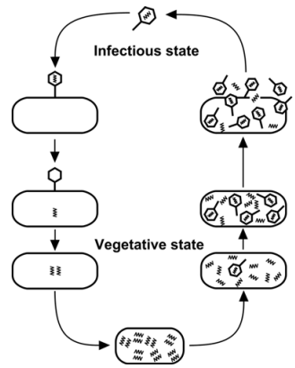
Figure 2–86. Cycle of phage
development (After Frey-Wyssling and
Mühlethaler, 1965).
The discovery of electron microscope in the end of third
decade of XX century made it possible to observe the viruses
(phages) visually. The first electron micrograph of a phage is made by
Ernst Ruska in 1940, and two years later — by S. Luria and T.
Anderson of T-even phages (T2 T4 and T6), parasitizing on E. coli.
Regarding the dimensions the phages were found to be very
small (about 0.1 nm, with a weight of 4×10⁻¹⁶ g), which is approximately one
thousandth compared to the bacterial host-cells. Their
development cycle is given in Figure 2–86.
The interest in viruses (particularly in phages) greatly increases.
especially after elaborating new methods of experimental analysis
and recording the recombinations and mutations in bacteria and
viruses (Luria, Delbrück, 1943; Luria, 1945; Delbrück, Bailey, 1946; Hershey,