juliana

Page 122

Figure 2–49. The hexoses fructose (A) and α- and β-glucose (B), main 
sources of chemical energy in the cells.

The system ADP⇆ATP is the carrier of chemical energy in these
processes. In the course of catabolic reactions an energy is released and
ADP-molecules can integrate a phosphate group thus forming ATP. When
running reactions are accompanied by energy consumption, the formed
ATP loses its end phosphate group and again is transformed into ADP (Fig.
2–50).

Figure 2–50. The system ADP⇆ATP, carrier of the chemical energy 
included in them. 

Monosaccharides, like proteins and nucleic acids, bind together and
form macromolecules. To a hydroxyl group of one monosaccharide an
aldehyde or keto group of another monosaccharide is joined with removal
of one water molecule. Thus, disaccharides, trisaccharides,
tetrasaccharides, etc. are formed. This process can continue by including
unlimited number of monosaccharides up to obtaining long-chain
polysaccharides containing hundreds or thousands monosaccharide
residues (Fig. 2–51).

Page 122 Read More »

Page 121

Depending on the number of carbon atoms they are trioses, tetroses,
pentoses, hexoses, heptoses and octoses. All monosaccharides, called
also simple sugars, are colourless crystal substances easily soluble in
water, most of them sweet to the taste.
Natural monosaccharides (with the exception of dihydroxyacetone)
possess optical activity expressed in turning polarized light to the right or to
the left. This phenomenon is due to the asymmetric carbon atoms in their
molecules. For example the triose glyceraldehyde has three carbon atoms,
and only one of them is asymmetric. It exists in two different stereoisomers
— D-right and L-left form (see Fig. 1–1). A hexose with four asymmetric
carbon atoms can have 16 isomers (2⁴=16).
The pentoses ribose and deoxyribose (Fig. 2–48) participate as
components in the nucleotides — the basic building block of the RNA and
DNA molecules. That determines their biological importance compared to
the other monosaccharides.

Figure 2–48. Structural formulas of the pentoses ribose and 
deoxyribose, participating in nucleic acids. 
A — β-D-ribose, a component of RNA; B — β-D-2-deoxyribose, a 
component of DNA.

Among the hexoses glucose and fructose (Fig. 2–49) are most spread
in nature. Both sugars have the same formula (C⁶H¹²O⁶), but the one is
aldose, and the other — ketose. Glucose is the main product of
photosynthesis. As a result of a number of consecutive redox processes
during the dark phase it is transformed into different carbohydrate
derivatives used to meet the energetic needs of the cells and in
synthesizing different organic components (see Chapter 1, Metabolism).

Page 121 Read More »

Page 120

These facts show that there are serious deviations from of the Watson
and Crick model in its initial form. The numerous unclear problems concerning
chromatin, the structural organization of the chromosomes and the different
genomes as a whole, the packing of the DNA-molecule by proteins, the
heterogeneous character of DNA and RNA with repeated nucleotide
sequences and satellite DNA, the reverse transcriptase, the differences in the
replication of the two chains, the interrupted gene structures, the mobile (IS)
elements, etc. render an opportunity for deep consideration. With the
postulation of the Central dogma in biology a transition from one extreme —
the leading role of proteins and environment, the inheritance of acquired
features in the process of development and vegetative hybridization for
changing hereditary features of organisms supported by T. D. Lysenko and his
followers with the denouncement of the objective regularities of Mendel,
Weissmann and Morgan laid the foundations of the chromosome theory, to
the other extreme — the positioning of DNA as fetish and placement of
proteins and RNA as secondary serving components. Where is the truth? The
servitude to dogmas is bad for science, especially biology. This problem must
be treated in the light of its evolutionary development and in this way to clarify
the origin, role and interactions between proteins and nucleic acids, otherwise
it would continue to give rise to “hot” discutions among the researchers in this
field.

Carbohydrates

Carbohydrates are the third basic component that participates in building
and functioning of unicellular and multicellular organisms. They constitute
about 80% of the dry mass of plant organisms and 2% of animal ones. In
contrast to nucleic acids and proteins, their biological role is less important
— they are sources of energy for realizing the metabolic processes and
components of cell membranes and tissues.
Sugars is a synonym of carbohydrates. They are: monosaccharides,
with a general formula (CH₂O)n, where n designates the number of C
atoms and can be every integer from three to eight; oligosaccharides,
containing from two to ten monosaccharide units connected with glucoside
bonds; polysaccharides — long chain built by a number of repeated
monosaccharide residues. Polysaccharide chains can be as linear, as well
as branched. Their molecular weight varies from a few thousand to 1
million.
Carbohydrates are built of only three chemical elements — carbon (C),
hydrogen (H) and oxygen (O). Each monosaccharide contains hydroxyl

Page 120 Read More »

Page 119

repeated and unique fractions there are intermediate fractions as well. The
specificity of genome organization in eukaryotes is reviewed in detail by
Ryskov (1989).
The repeated nucleotide sequences were discovered by Britten and
Kohne (1968). Though to a great extent arbitrarily, they are subdivided
into manifold and moderately repeated ones. It is believed that the
intermediate fraction consists of a number of families, one of which being
the satellite DNA. Their biological role and importance in the
organization of the genome remain unclear.
The transfer of genetic information from RNA to DNA due to the
reverse transcriptase proved a sensational interest. Its experimental
proof started with the publications of Temin (1960, 1964 a, b) on some
oncogenic viruses. In only a few years time it was confirmed by other
authors (Spiegelman et al., 1965, 1970 a, b; Baltimore, 1970; Temin,
Mizutani, 1970; Temin, Baltimore, 1972, etc.).
In the beginning the reverse transcriptase was met with a wide
distrust. It was considered impossible, erroneous or an isolated case in
certain oncogenic viruses. After that the scope of the studies was
broadened and it was confirmed on non-oncogenic viruses, cancer
tissues and intact cells including cells of higher organisms. At present
reverse transcriptase is well-established fact in science.
Normile’s report (1996) on the hypothesis of the Japanese scientists
M. Furusawa and H. Doi is now arousing similar surge of interest. They
have suppose that both DNA chains replicate separately from one
another. The leading chain is constantly synthesized in the direction in
which the helix is uncoiled. The other chain — the delayed one — is
synthesized in numerous small fragments in the opposite direction. The
two processes engage different enzymes and differ in the mechanism of
error correction. This duplicitous procedure gives rise to more errors in
the delayed chain which has made Furusawa assumed certain
evolutionary consequences involved.
A multitude of other established data and facts related to the
structural organization and functions of the genome cannot be under
estimated and ignored. These are the multigenic organization of the
genes in eukaryotic cells, which leads to the increase of their numbers
and the entire genome; the interrupted structure of the genes, as a result
of which a lot of genomes are organized as individual transcription units;
the switch on and off of introns and exons, the binding of which is
accomplished by RNA splicing; the control elemets of McClintock (1950,
1956 a, b) called mobile IS-elements (insertion sequences) capable of
breaking away and inserting themselves in new genome sites, etc. They
are believed to exercise influences on the normal activity of the genes,
the state of the genome and its stability.

Page 119 Read More »

Page 118

image

Figure 2–47. Minor, i.e. rare pyrimidine (A) and purine (B) bases found in 
DNA.

The study of the kinetics of DNA renaturation in different organisms
has shown that in the different fractions it takes place in inverse proportion
to the complexity of the sequences. The greater rate of renaturation of the
individual fraction is due to the fact that it consists of nucleotide sequences
that are multiply repeated. When the renaturation process takes place at a
lower rate, the nucleotide sequences are assumed to be unique or
unrepeated. Literature data show that the sizes of the fractions with
repeated sequences can differ considerably — from 5 to 80% (an average
of 30—40%), as about 50 to 60% of the genome of the studied organisms
consisting of repeated nucleotide sequences of an average length of 300
nucleotide pairs that are intermittently replaced by unique sequences of a
length of 700 to 1100 nucleotides (see Hadjiolov et al., 1976). Except the

Page 118 Read More »

Page 117

i.e. rare bases — 5-methylcytosine, 5-hydroxymethylcytosine, 2-methyl-
aminopurine, 6-methylaminopurine, etc. (Fig. 2–47) were discovered.
These bases lead to errors in the binding to other bases and exert an
influence on the replication and translation processes. Minor bases are also
found in RNA.

The road to the heterogeneous character of DNA is open. The
assumption that the eukaryotic cells contain much more genetic material
than it is needed according to the calculations made and the absence of
correlation between the complexity of a given organism and the size of
its genome was confirmed experimentally. Using the methods of
denaturation, hybridization and reassociation of DNA, the presence of
unique and reoeated nucleotide sequences was established as well as
the so-called satellite DNA.
For the first time a satellite DNA-component was observed by Kit
(1961) upon centrifugation of mouse DNA in neutral density gradient of
CsCl. Kit has established that about 10% of it is found in the light band
(p=1.691 g.cm³) compared to the major zone (p=1.700 g.cm³), which is
due to the decreased G—C content. A characteristic feature of this
fraction is that it differs in its nucleotide composition from the rest of the
major part of DNA. Satellite components are also found in many other
eukaryotic cells. In some species they comprise more than 30% of the
total amount of nuclear DNA, varying in nucleotide composition.

Page 117 Read More »

Page 116

contrast to heterochromatin the euchromatin is despiralized during the
interphase, it is evenly distributed in the nucleus and is more poorly stained.
The euchromatin portions are thought genetically active since it is believed
that genes are predominantly concentrated in them, while the
heterochromatin is genetically inactive. Since this problem is unclear, there is
no ground to think that heterochromatin is an inert mass devoid of any
biological importance.

image

Figure 2–46. Two differentially stained 
metaphase chromosomes of barley (Hordeum 
vulgaris). The dark areas indicate to the 
 portions of structural heterochromatin and the 
light ones — to euchromatin (Courtesy of K. 
Gechev, Institute of Genetic Engineering, 
Kostinbrod). 

Together with the long lived idea of chromatin as a smooth deoxynucleoprotein
fibres (DNP-fibre) of more or less regular super-spiralization, other models have been
launched according to which DNA is coiled in or around protein complexes localized
along the DNP-fibre. Olins and Olins (1974) have established that chromatin has a granular
structure. According to them chromatin fibres are built of bound spherical bodies of a
7—10 nm diameter (called nucleosomes) which are localized along the chromatin
fibres in a bead-like fashion. This model has aroused a great interest.

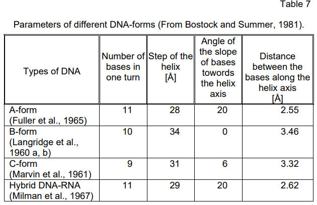
Some Deviations from the Watson and Crick Model

The model of the DNA double helix forwarded by Watson and Crick
corresponds to a configuration of the B-form type with a right turn. Further
investigations (Langridge et al., 1960 a, b; Marvin at al., 1961; Fuller et al.,
1965, etc.) have established that DNA can exist in three different
configurations — A-, B- and C-forms which under definite conditions can
convert into one another. The major parameters of these DNA-forms are
presented in Table 7. According to Davies and Zimmerman (1988). DNA in
chromatin assumes a special Z-conformation. Lejeune (1979) has
established a left rotation of DNA. These data are quite indicative of
nature’s capacities to create a variety of forms of existence even of such
strictly specific high-molecular compounds as DNA.
Besides, it was proved that apart from the purine/pyrimidine ratio the
ratios of the incorporated in their composition DNA-bases A : T and G : C = 1
varies to a great extent in the DNA of higher plants and animals. In many
cases A + T > G + C. Such a DNA was called the AT-type. Also the minor,

Page 116 Read More »

Page 115

themselves in the replication of the chromosome in a non-random fashion, thus
being handed down from one cell generation into another (just like DNA), which
is in accordance with the idea of Tsanev and Sendov (1971) that the specific
arrangements of histones along the DNA-chain are kept up during the replication
by specific interactions between the previously existing DNA-bound histones
and the newly-synthesized free histones. Some authors would not agree with
this assertion (Jackson et al., 1975, 1976; Seale, 1976). According to them
histones are randomly distributed upon the self-replicating chromosomes. This is
a serious scientific disagreement. The importance of this problem requires a
definitive solution, which is related to further investigations. More plausible,
however, seems from a general biology standpoint the thesis supporting the
non-random i.e. conservative distribution of histones on both DNA-chains.
The established by Chentsov and Poljakov (1970) presence of an
especially dense substance in the metaphase and telophase chromosomes
distinguished for its density from the surrounding cytoplasm and chromatin,
called matrix deserves no less attention (Fig. 2–45). According to Baskin
(1995) nuclear matrix is the key to gene expression.

image

Figure 2–45. Matrix in metaphase and telophase chromosomes (After 
Chentsov and Poljakov, 1970). 
a — metaphase chromosome after processing the cells with 0.2% of 
CoCl₂ solution; b — telophase chromosome surrounded by matrix. 
M — matrix

By the help of the methods for differential staining of chromosomes the
existence of two fractions of chromatin — heterochromatin and euchromatin
— was proven (Fig. 2–46). They differ in a number of qualities and
properties. Heterochromatin remains condensed during the entire interphase
and preserves its capacity for an intense staining, which is typical of
metaphase chromosomes. Brown (1966) has arrived at the conclusion about
the existence of two heterochromatin types — structural and facultative. In

Page 115 Read More »

Page 114

image

Figure 2–43. A scheme illustrating 
 the semi-conservative manner of 
DNA- replication. Each of the two 
 chains serves as a matrix for the 
formation of a new complementary 
chain (After Meselson and Stahl, 
1958). I — initial (parental) molecule; II — 
first generation (“daughter” 
molecules); III — second generation 
(molecules — “grandsons”)

image

Figure 2–44. Schematic presentation of 
 the DNA-replication. The parental chains 
separate at the replication fork. Each 
parental chain serves as a template for 
the synthesis of a new chain.

The packing of DNA with proteins is a key problem. That is why it provokes general
interest. In their monograph “The Eukaryotic Chromosome” Bostock and
Sumner (1981) pay attention to two unsolved questions related to the linear
organization of the nucleosome structures: Whether DNA is found outside the histone
complexes or is it situated inside them? Is the entire DNA in the nucleus packed in one
and the same fashion or are there separate portions in the chromatin (transcribing or
replicating chromatin) that are packed in another way? Of a great interest is the
publication of Tsanev and Russev (1974) on the distribution of the newly-synthesized
histones at the time of DNA replication. The authors have proved experimentally that during the
replication the existing histones remain bound to the parental DNA-chain,
while the newly-synthesized histones bind to the new DNA-chain.
The results obtained by them confirm the ones of Hanckok (1969, 1970)
postulating that histones are conservative elements of chromatin distributing

Page 114 Read More »

Page 113

It is known that except for DNA a number of different proteins are present
in the chromosomes of eukaryotic cells. The complex of nucleic acids and
proteins is called chromatin. According to data of Hadjiolov et al. (1976)
chromatin isolated from interphase cells after the separation of cytoplasmic and
soluble nuclear components usually contains about 30—40% DNA, 3—5%
RNA and about 50—60% proteins, and according to Lehninger (1976) human
metaphase chromosomes contain roughly 15% DNA, 10% RNA and 75%
protein. These data about the high percentage of protein content in the
chromosomes should not be ignored. DNA-molecules in prokaryotic cells,
viruses including, are also bound or packed with proteins.
The presence of RNA in the composition of chromatin is not
sufficient ground for many researchers to accept it as an integral component of
the chromosomes. It is assumed that RNA is a product of DNA transcription.
This standpoint is contradictory to the opinion of certain authors presuming that
RNA precedes DNA as genetic material (see Lazcano, 1986).

The proteins taking part in the chromatin composition are divided into
two groups: histones and non-histones. The first comprise a group of low
molecular proteins rich in lysine and arginine denoted as H1, H2A, H2B,
H3, H4, H5. The last one (H5) is found in the erythrocytes when they have
nuclei. In the spermatozoa of some fish, amphibia and other animal species
histones are replaced by protamines. The so-called acidic proteins which
are very diverse in composition and possess totally different structures and
properties are classified as comprising the second group. This group is the
least studied one.

image

Figure 2–42. Electron micrograph
of native portion of the DNA-molecule (After Stahl, 1966).

Page 113 Read More »