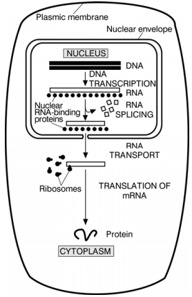
juliana

Page 112

image

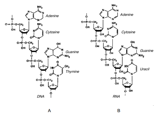
Figure 2–41. Three-dimensional models of the double helices of DNA (A) 
and RNA (B). Both structures are formed from two antiparallel 
polynucleotide chains linked by hydrogen bonds between the 
complementary bases. DNA is presented in B-form, and RNA — in its A 
form (Courtesy of Sung-How Kim; From Alberts et al. 1986). 

The Watson and Crick model places the DNA molecule in a central
position and the protein components in a subordinate one. Since this is
one of the key issues of the modern biology and genetics it cannot be
left out without the due attention.

Page 112 Read More »

Page 111

Figure 2–40. The X-ray diagram of the B-form of DNA (After Franklin and Gosling, 1953).
This diagram has played a definitive role in revealing the spatial structure of DNA. The
cross-like dark reflections prove that the structure is helical. The heavy black regions at the top and
bottom show that the purine and pyrimidine bases (3.4 nm thick) are regularly stacked next to
each other, perpendicular to the helical axis.

image

The three-dimensional models of the double DNA and RNA helices are 
shown in Figure 2–41 and the electron micrograph of a native portion of the 
DNA-molecule — in Figure 2–42. 

With the discovery of the DNA double helix and the possibilities for
replication which such a helix renders, a lot of the problems of biology
and cytology were solved. It became clear what the chromosome
behaviour in the nucleus after the fertilization was due to, as well as the
nuclear division and observed even distribution in the process of
division. The phenomena of transformation, transduction and conjugation
which represent the transfer of hereditary material (DNA) from a donor
cell to a recipient cell found their explanation. The studies in that field
were spread on a large scale. A number of microorganisms became the
model objects for such a kind of study, such as bacteria, viruses,
microalgae, fungi, etc.
An important question arised: how the DNA replication is realized?
The answer was provided by Meselson and Stahl (1958). In experiments
with E. coli bacterial cultures using the method of the labelled atoms (¹⁴N
and ¹⁵N) and centrifugation at a dense gradient of CsCl they have
established that DNA-replication is taking place in a semi-conservative
fashion (Fig. 2–43). This coincides with the hypothesis of Watson and
Crick, according to which the two initial complementary DNA-chains are
uncoiled and each serves as a template on which the new complementary
chain is synthesized (Fig. 2–44).

Page 111 Read More »

Page 110

were replaced by DNA. Possessed with the idea of immortality of the
genes Watson put on the wall behind his desk a sheet of paper with the
inscription: “DNA→RNA→Protein”, which turned into the Central dogma
of molecular biology (see Crick, 1970).

image

Figure 2–39. DNA double helix (After Alberts at al., 1989).

A tribute to the revealing of the spatial structure of DNA have also
other researchers whose results have prepared the terrain for this
discovery accepted as the greatest in biology of the XX century: the first
X-ray studies on nucleic acids (Astbury, Bell, 1938; Astbury, 1947); the
Pauling and co-authors models of the helical configuration of the
polypeptide chains and nucleic acids (Pauling, Corey, 1951 a—c, 1953;
Pauling et al., 1951 d); the X-ray studies of Wilkins at al. (1953 a, b) on
the spatial structure of DNA; the famous X-ray diagram of Rosalind
Franklin and Gosling (1953) — Fig. 2–40 that has experimentally proved
the suggestions of the helical structure of DNA, and E. Chargaff’s rules
about the equimolar quantities of the purine and pyrimidine bases
(Chargaff, 1950).

Page 110 Read More »

Page 109

Figure 2–38. Portions of the polynucleotide chains of DNA (A) and RNA (B). 

The Watson and Crick Model

The authors of the model — James Watson and Francis Crick (1953 a)
have established that the DNA molecule is a double helix consisting of
two complementary chains built from four bases — adenine (A), guanine
(G), thymine (T) and cytosine (C). The bases are situated inside the helix
linked by hydrogen bonds, and the sugar-phosphate skeletons are
outside forming the frame. At that, adenine is always bound to thymine
(A—T) and guanine to cytosine (G—C). The bound bases lie on planes
perpendicular to a helix axis and are located one under the other at a
distance of 0.34 nm. A full turn is accomplished by 10 pairs of bases
forming a step of 3.4 nm (Fig. 2–39).
This model has fully satisfied the requirements posed by the then
existing data from the X-ray diffraction studies. Besides it revealed the
possibility of self-replication, since the DNA molecule comprises two
complementary chains which can be uncoiled and each of the initial chains
may serve as a template for the synthesis of a new complementary chain
(Watson, Crick, 1953 b).
The biological essence born by this discovery was so great that it
has brought its authors immediate recognition and gave a strong impetus
to the development of biology, molecular biology and genetics in
particular. Proteins regarded as eventual bearers of heredity until then

Page 109 Read More »

Page 108

Figure 2–37. Nitrogenous bases and sugar components participating in the 
DNA and RNA build-up (A) and nucleotides of DNA and RNA (B).

Page 108 Read More »

Page 107

life processes (see the last paragraph of Section 2. 6). That is probably
why the number of enzymes is incredibly great, so that they can carry
out their numerous and various functions.
This assumption raises the question whether a bacterial cell (e.g. E.
coli
), if it is to perform exactly the scheme prescribed to it by man for the
protein synthesis, would be able under optimum conditions to complete
its development cycle and divide for 20 or 30 min which is in fact easily
accomplished. The doubts in this respect are huge. That is why the
opinion expressed by Alberts et al. (1986) is considered true and is
gladly shared: “…Molecular processes underlying protein synthesis are
exceptionally complicated and their study has only been accumulation of
facts for the time being, without encompassed in a comprehensive
theory. The discovery of the mechanisms involved in protein synthesis
could throw some light as well on those early events related to the origin
of life itself”. Besides, the authors accepted that from a chemical point of
view proteins are “the most complex of all molecules known”.
The problem of protein self-replication without the participation of
DNA remains unsolved. This is a heresy since it contradicts the Central
dogma in molecular biology, accepting only DNA as the unique molecule
endowed with such a capacity. A great interest is provoked by the data
of Lee et al. (1996) on the existence of self-replicating peptide. In the
literature there are also other studies in that trend. If it is confirmed more
convincingly, it will be important for the throwing light on protein
synthesis especially in the early stages of the evolution of living matter
and cell structures.

Nucleic Acids

As a result of the prolonged studies on nuclein two types of nucleic acids
were discovered — deoxyribonucleic acid (DNA) and ribonucleic acid
(RNA). They proved to be long polynucleotide chains of great biological
importance. They are built from four bases each, which in combination
with one of the pentose sugars and phosphate residues form the
corresponding nucleotides (Fig. 2–37). The differences between them are
expressed in the following: DNA contains deoxyribose while RNA
contains ribose; thymine in DNA is replaced by uracil in RNA. The binding
of the nucleotides leads to the formation of long polynucleotide chains of
DNA and RNA (Fig. 2–38).
The biological role of DNA was made clear after the discovery of its
spatial structure. The history of this great discovery is very thrillingly
described in J. Watson’s book “The Double Helix” (1968, 1975).

Page 107 Read More »

Page 106

accomplished by another specific group of enzymes — aminoacyl-tRNA-
synthetase
leading to the formation of an aminoacyl-tRNA-molecule. For
each amino acid there is a separate aminoacyl-tRNA-synthetase: (one of them
joins glycine to — tRNAᵍˡʸ another — alanine to — tRNAᵃˡᵃ, etc. The extension of
the polypeptide chain is continued until one of the three stop-codons is
reached, which is a signal for stopping the synthesis.

Figure 2–36. Scheme (A) and spatial model (B) of codon and anticodon in 
the molecule of the phenylalanine tRNA of yeast (Courtesy of Sung-Hou 
Kim; From Alberts et al., 1986)

It is supposed that in prokaryotic cells transcription and protein
synthesis take place almost simultaneously. Here the main unsolved
problem is how the despiralization of the circular DNA-molecule is
effectuated at the time of the replication. Most probably this occurs by its
severing at a given segment under the action of one or more enzymes.
These processes are not clearly elucidated.
One of the most important functions attributed to proteins for now is
the specific catalysis of biochemical reactions, i.e. their action as
enzymes. This differs them a lot from nucleic acids. It is considered that
oxygen transfer is a characteristic property of hemoglobin and not of the
gene encoding for that protein. It is quite possible from such a standpoint
to draw the conclusion that although DNA contains the whole genetic
information necessary for the structure and development of the cells, it is
kept in an “academic” state and does not immediately participate in the

Page 106 Read More »

Page 105

When the first DNA polymerase (DNA-polymerase I) was discovered, the
transcription mechanism of the DNA chain appeared to be comparative simple.
Afterwards it proved to be much more complex. In the small bacterial cell E. coli
there have been identified not one but three DNA-polymerases — I, II and III.
For some time it was thought that DNA-polymerase I is the main polymerizing
enzyme binding the deoxynucleotides. It is now accepted that this role is played
by DNA-polymerase III, i.e. the last one in the order of its discovery. The function
of DNA-polymerase II remains unclear.
In eukaryotic cells three forms of RNA-polymerase with different functions
have been identified as well. One of them is responsible for the rRNA
synthesis, the other one — for the mRNA and the third one — for tRNA.
It is accepted in principle that any DNA-segment can be transcribed
with the formation of two different mRNA molecules (one for each of the
two DNA-chains). In reality, it is more likely for this to take place on only
one of them, which is determined by the promotor (start-signal) in DNA.

Figure 2–35. A scheme clarifying the 
transition of mRNA from the nucleus 
into the cytoplasm where by the help of 
the ribosomes the translation and 
synthesis of polypeptide chains 
(proteins) take place in eukaryotic 
cells


The mRNA synthesized in the nucleus of eukaryotic cells passes
into the cytoplasm and binds to the ribosomes. There the process of
translation of mRNA genetic information into polypeptide chains is
performed (Fig. 2–35). This process is very complex and multistage, since
translation is not directly connected with the α-amino acids which are to
be polymerized. The preparation of their incorporation into polypeptide
chains is started with the amino acid COOH-group activation by ATP and
its transfer on a specific transport RNA (tRNA). For each of the 20
amino acids participating in protein synthesis there are one or more
tRNAs. An important role for the successful decoding of the information
in mRNA is played by the precise binding of the bases between the
mRNA codon and the tRNA anticodon (Fig. 2–36). Each amino acid is
attached to the corresponding tRNA molecule specific for it. The
“recognition” of the amino acid by tRNA and its adaptation to it is

Page 105 Read More »

Page 104

is formed growing in the direction from 5′ to 3′ end with the incorporation of
nucleotides. This process goes on until another specific nucleotide sequence
is reached — stop-signal i.e. the signal for termination. Then the RNA
polymerase is separated from the DNA-chain matrix and the newly
synthesized mRNA. The hybrid helix RNA—DNA thus formed is unstable, that
is why the initial DNA-helix is restored and the completed RNA chain is
released as a free single-chain molecule (Fig. 2–34).

Figure 2–34. Scheme illustrating how RNA polymerase initiated 
synthesis at the special start signal in DNA, called promotor, and 
terminates at the stop signal, after which the enzyme and the 
synthesized new mRNA chain are released. The rate of polymerization 
at 37°C equals 30 nucleotides in a second. The picture gives an idea of 
the approximate ratio between the sizes of RNA polymerase and the 
DNA helix (After Alberts et al., 1989).

Page 104 Read More »

Page 103

image

Figure 2–33 B. Quaternary structure of the hemoglobin molecule as 
determined by X-ray analyses (Based on Kendrew, 1961, 1963; Perutz, 
1962, 1963; From Stent, 1974)

The problem of protein synthesis represents a “hot point” in biology
especially in its evolutionary aspect (see Chapter 1, Section 1. 6). Since
there are still no convincing data about self-replicating spiralized or non-spiralized
polypeptide chains this issue will be treated in the light of the
presently available conventional concepts.
According to modern views genetic information is encoded in the DNA
molecule. Under the action of the enzyme RNA-polymerase the process of
transcription of DNA is accomplished with the formation of messenger RNA
(mRNA). Transcription starts with RNA-polymerase joining a specific
nucleotide sequence in DNA called promotor. The double helix of DNA is
despiralized and at a definite portion of it a complementary RNA—DNA chain

Page 103 Read More »