juliana

Page 162

Reparation Processes

The primary damages and changes occurred in the genetic material are
only a stage of the mutation process. Not less important is the next stage
connected with the further “fate” through the “laboratory labyrinths” of cell
structures determining the heredity. Practice and experiments show that not
all of them are firmly included in the genetic material and complete with
mutations. Obviously, there exist mechanisms which repare or remove
them in some way, thus maintaining the normal status (status quo) of
organisms. These processes have been called reparation.
If bacterial cells are subjected to the action of high doses of UV-radiation
(~280 nm), their ability to form colonies abruptly decreases. It has been
observed that in some bacteria this ability is restored after their exposure to
daylight, that has led to the discovery of phenomenon photoreparation or
photoreactivation. For the first time it is observed by Kelner (1949 a, b) at
lighting up Actinomyces suspension, but it is confirmed on a number of other
microorganisms — bacteria, phages, paramecia, etc.
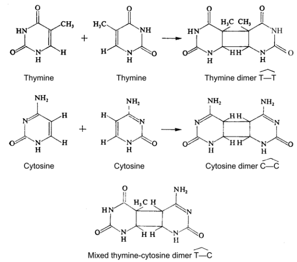
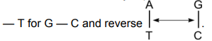
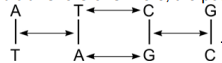
It has been established that by UV-irradiating dimers of thymine (see
Fig. 2–80), are formed in DNA, which disturbs the structure and functions of
the genes. Except between thymine bases (TT), dimers may occur between
uracil and cytosine (UC) and only between cytosine (CC). The more are the
dimers formed, the greater is the lethal effect. After photoreactivation the
dimers disappear (Setlow. Setlow. 1962).
The beginning of more profound studies on the processes of reparation
is laid by revealing the enzyme photoreactivation (Setlow, Setlow, 1963) and
elucidating the mechanism of so-called dark reparation of the damages in
DNA. Many bacteria repair the damages caused by UV-rays in dark. That has
led to the supposition of existence of different reparation mechanisms. Some
bacteria have shown greater susceptibility to radiation, other have been
found more resistant. It proved that during dark reparation without any
assistance the resistant lines remove pyrimidine dimers in DNA, while the
susceptible ones do not remove them (Setlow, Carrier, 1964; Boyce, Howard
Flanders, 1964; Howard-Flanders, 1968. 1973). According to Auerbach
(1976) photoreparation can achieve 100% effectiveness and correctness of
the transformation of dimers into monomers.
On the basis of performed investigations, the mechanisms of
reparation are reduced to three types: a) photoreactivation; b) reparation
through cutting (excision reparation); c) postreplication reparation.
Photoreactivation is the most simple mechanism, since only one
enzyme is required that should be capable to “recognize” and bind the part
of DNA (thymidine dimer) underwent a primary damage. The source of
energy is visible light, which serves the photoreactivating enzyme in
separating the dimer and restoring the initial state. It is established that
daylight is most effective between 310 and 440 nm (Setlow. 1966).

Page 162 Read More »

Page 161

Figure 2–82. Basic skeleton in the molecules of N-nitrosocontaining 
supermutagens (A) and the radical of Gordy and co-authors (B) 
determined by EPR-spectroscopy.

According to Bostock and Sumner (1981) it is undoubtedly proven
that in mammals chromosome damages arise as a result of the action of
radioactive irradiations on proteins. They give reasons for their opinion on
the basis of the results of other authors showing that in the cells of
chinese hamster the lethal effect and chromosome aberrations are
caused by irradiations, whose specter is different from the absorption
specter of DNA (Chu. 1965; Rauth. 1970) and resemble that of tyrosine
containing proteins (Zirkle. Uretz. 1963).

Figure 2–83. Diagram illustrating intercalation of
acridine dyes in DNA-molecule (After L. S. Lerman, 1964;
From Bostock and Sumner, 1981).

Some chemical compounds play the role of mutagens. without causing direct
damages in DNA. These are so-called intercalated agents. Among them.
proflavin, acriflavin, and acridine orange are better studied.
Lerman (1961. 1963) has shown that acridinic dyes form complexes with DNA. At
that their planar molecules penetrate (intercalate) between neighbouring
nucleotide bases, which leads to increasing the distance between them (Fig. 2–83). It is
supposed that acridine molecules, after eir interaction with DNA, induce incorrect
complementation of the nucleotide bases and mutations with shifting the frame of reading.

Page 161 Read More »

Page 159

After experiments carried out on the unicellular green alga
Scenedesmus acutus (Nicolov, 1973 a—c, 1975) it was established that for
the extremely high mutagenic effect of N-nitrosocontaining supermutagens,
except an alkyl radical, is necessary also the presence of a highly polarized
and unbroken

bond in combination with the radical.

Figure 2–81. The structural formula of ethyl-N-nitrososarcosine 

A reason for such a conclusion is the absence of mutagenic effect in ethyl-
N-nitrososarcosine. As it is seen from its chemical formula (Fig. 2–81), this
compound differs from N-nitroso N methylurethane and N-nitroso N ethylurethane
(see Table 9) in that the ─ N ─ C ⎯ bond is interrupted by one methylene group (⎯CH₂⎯).
For that reason the mutagenic effect of ethyl-N-nitrososarcosine is 0.02—0.25%, i.e. within
the borders of spontaneous mutations, compared to 38.59% for N-nitroso N-ethylurethane
and 57.10—100% for N-nitroso N-methylurethane (Table 10).
Weak mutagenic effect was manifested by N-nitrosodialkylamines
which doubted their belonging to the group of supermutagens. By
comparing their chemical formulas with those of N-nitrosoalkylurethanes,
N-nitrosoalkylureas and N-nitroso-N′-nitroalkylguanidines (see Table 9) it is
obvious that, though possessing not one but two alkyl radicals, N-
nitrosodialkylamines represent peculiar fragments of the hypothetic
monopeptide units, which are the basic skeleton in the other three groups.
That was the reason to accept that breaking the integrity of
monopeptide ion, which is the basic skeleton in the molecules of N-
nitroso-containing supermutagens, is the cause for the absence of
statistically reliable mutagenic effect of ethyl-N-nitrososarcosine and of the
homologous series of N-nitrosodialkylamine group (Nicolov, 1973 a, b).
Comparing the basic skeleton in the molecules of N-nitroso-containing
supermutagens with the radical, established by Gordy and coauthors
(Gordy, Schields, 1960; Patten, Gordy, 1960) after treating native proteins
with ionizing radiations and determined by EPR-spectroscopy, one is
impressed by the great similarity between their structures (Fig. 2-82). The
obtaining relatively stable radicals after irradiation of proteins having a
structure similar to that of the main skeleton of N-nitroso-containing
supermutagens suggests that it is possible to exist some community of the
mechanisms connected with the initial stages of physical and chemical
mutagenesis, their further action being realized by genetically-active
structures of the cells (Nicolov, 1973 a).

Page 159 Read More »

Page 157

Figure 2–80. Pyrimidine dimers. 

The mutation changes in chromosomes can be highly varied — from
changes in nucleotide composition and mistakes in binding the bases in
DNA to great structural transformations in them. These changes are
caused by different reasons, a lot of them being unclear. Some of them are
the already mentioned ionizing radiations and chemical mutagens. Among
the latter a special place is held by the alkylating compounds, some of them
known as supermutagens (Table 9).
The mechanisms of action of alkylating compounds are complex. They
cause various genetic changes — transitions, transversions, chromosome
aberrations, etc. It is accepted that these substances express their
mutagenic effect in two different ways. The one is a direct formation of
“incorrect” nucleotide pairs, and the other — alkylation of DNA-bases at
different positions through free radicals, like methyl (⎯CH₃), ethyl (⎯C₂H₅),
propyl (⎯C₃H₇), etc. In vitro alkylation has shown a preference to guanine
in 7th position (Lawley, 1966), while in vivo — great is the role of oxygen
atoms in 4th position for thymine and in the 6th for guanine (Auerbach,
1976; Singer, 1976).

Page 157 Read More »

Page 156

Figure 2–79. Deamination of the usual bases in DNA and formation of 
unusual ones, leading to disturbances in the correct complementation. 

Ultraviolet radiation in the range ~ 260 nm (i.e. wavelength
corresponding to the maximum absorption by DNA) also causes different
alterations in nucleic acids. Among them well-known is the formation of
pyrimidine (mainly thymine) dimers (Fig. 2–80). If they prove to be stable,
they may have an adverse effect on DNA-ability to replicate.
The majority of the mutations are not base substitutions. They
represent large deletions or changes caused by a shifting the frame of
reading (frame shift mutations). The latter are due to an addition or loss of a
certain number of nucleotides in DNA-chain, that leads to mistakes in the
normal reading of codons and therefore — to defects in the translation of
genetic information. Thus, during protein synthesis the correct amino acid
sequence is disturbed.
Except the point mutations, the chromosome and genome mutations
are of great interest. Their molecular bases and mechanisms are less
studied. They are connected with changes in the structure and number of
chromosomes as well as with the ploidy of chromosome set.

Page 156 Read More »

Page 155

Figure 2–78. Properties of 5-bromouracil (a) and 2-aminopurine (b) 
relevant to the formation of “incorrect” nucleotide pairs with the bases of 
DNA. Hydrogen atoms are denoted by black circles, and the bonds 
between them — by dotted line (After Hayes, 1965). 

Under the action of different chemical mutagen factors the bases of
DNA can undergo a modification that leads to inducing of mutations. For
example, nitrous acid (HNO₂) causes an oxidative deamination of the
bases, at that amino groups are substituted for keto groups. Thus, cytosine
is transformed into uracil, adenine — into hypoxantine, and guanine — into
xantine. Thymine is the only, that is not a subject to deamination since it
lacks amino group, and contains a keto group (Fig. 2–79). At that uracil
binds adenine, and xantine and hypoxantine — cytosine.

Page 155 Read More »

Page 154

by some analogues of pyrimidine and purine bases. These analogues
possess a structure very similar to that of usual bases — adenine, guanine,
cytosine and thymine. 5-bromouracil and 2-aminopurine are the best
studied among them.

Figure 2–77. “Prohibited” nucleotide pairs, which arise when some base 
is in a rarely met tautomeric form. On the left — bases underwent the 
tautomeric transition. On the right — bases in the form usually met in 
DNA (After Rukmansky et al., 1984).

5-bromouracil (BU) is an analogue of thymine, in which the 5-
position methyl group (—CH) is substituted for brom atom (Br).
Normally, thymine and BU (in keto-form) bind adenine (T—A or A—BU).
When BU is in the rarely met enol-form, it forms hydrogen bonds with
guanine (G—BU). 2-aminopurine (AP) is an analogue of adenine and
should form a nucleotide pair with thymine (T—AP). If it is in imino-form,
it binds cytosine (C—AP) — Figure 2–78. Incorporating the base
analogues into the molecule of DNA leads to mistakes in
complementation with other bases which, on its part, causes replication
mistakes because of the realized transition AT ⇄ GC.

Page 154 Read More »

Page 153

Mutations can be forward and reverse. Forward mutations represent
lead to hereditary changes in the genes of naturally existing species, and
the reverse — to restoring the initial form which can be full or partial. If the
mutations are connected with a loss of genetic material, then a reverse
mutation is impossible.
Also, there exist other classifications of the mutations — generative
and somatic, dominant and recessive, vital and lethal, fertile and sterile,
etc. They have a rather formal character.
Molecular Bases and Mechanisms of the Mutation Changes
This problem is the essence of the mutation process. That is why it attracts
the attention of many investigators. Its elucidation will answer to a number
of questions about the mechanisms of realizing the changes in living
organisms.
Though suddenly arising, the mutation changes are linked with some
processes that prepare their appearance. Undoubtedly these processes are
related to the hereditary material, since the mutations are inherited. The data
show that a considerable part of the mutations are a result of changes in the
base sequence of DNA-molecule. Most of them are obtained as a result of
investigations on phages and bacteria, but it is considered that their
mechanisms are universal in the living nature. For the present, best studied
are the molecular processes leading to the appearance of point mutations.
That is why, they will be considered in more detail.
Point mutations, as it was already mentioned, are due to changes of
single nucleotides in the molecule of DNA. The theoretical analysis of Frees
(1959 a, b) has shown that two types of nucleotide substitutions can exist.
In one of them there is realized a substitution of pyrimidine for another
pyrimidine or of purine for another purine. This type of substitutions are
called transitions. They represent a substitutions of the nucleotide pair A

In the other type of nucleotide substitutions a pyrimidine is substituted
for a purine, or a purine is substituted for pyrimidine. This type of
substitutions are called transversions. Here, the possible substitutions are

These substitutions can lead to arising of so-called “prohibited” or
“wrong” nucleotide base pairs. If some base is in a rarely met tautomeric
form, it can make “incorrect” hydrogen bonds with another base: T—G,
C—A, A—C and G—T (Fig. 2–77).
The appearance of “incorrectly” bound base pairs in DNA that leading
to replication and mutation changes of the genetic material, can be caused

Page 153 Read More »